Abstact
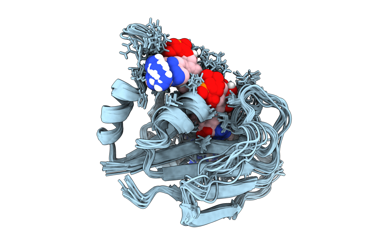
There is a significant need for new therapeutics to treat infections caused by the biodefense agent Bacillus anthracis. In pursuit of drug discovery against this organism, we have developed novel propargyl-linked inhibitors that target the essential enzyme dihydrofolate reductase (DHFR) from B. anthracis. Previously, we reported an initial series of these inhibitors and a high-resolution crystal structure of the ternary complex of the enzyme bound to its cofactor and one of the most potent inhibitors, UCP120B [Beierlein, J., Frey, K., Bolstad, D., Pelphrey, P., Joska, T., Smith, A., Priestley, N., Wright, D., and Anderson, A. (2008) J. Med. Chem. 51, 7532-7540]. Herein, we describe a three-dimensional solution structure of the ternary complex as determined by NMR. A comparison of this solution structure to the crystal structure reveals a general conservation of the DHFR fold and cofactor interactions as well as differences in the location of an active site helix and specific ligand interactions. In addition to data for the fully assigned ternary complex, data for the binary (enzyme-cofactor) complex were collected, providing chemical shift comparisons and revealing perturbations in residues that accommodate ligand binding. Dynamics of the protein, measured using (15)N T(1) and T(2) relaxation times and {(1)H}-(15)N heteronuclear NOEs, reveal residue flexibility at the active site that explains enzyme inhibition and structure-activity relationships for two different series of these propargyl-linked inhibitors. The information obtained from the solution structure regarding active site flexibility will be especially valuable in the design of inhibitors with increased potency.