2KFG image
Deposition Date 2009-02-20
Release Date 2009-12-22
Last Version Date 2024-05-01
Entry Detail
PDB ID:
2KFG
Keywords:
Title:
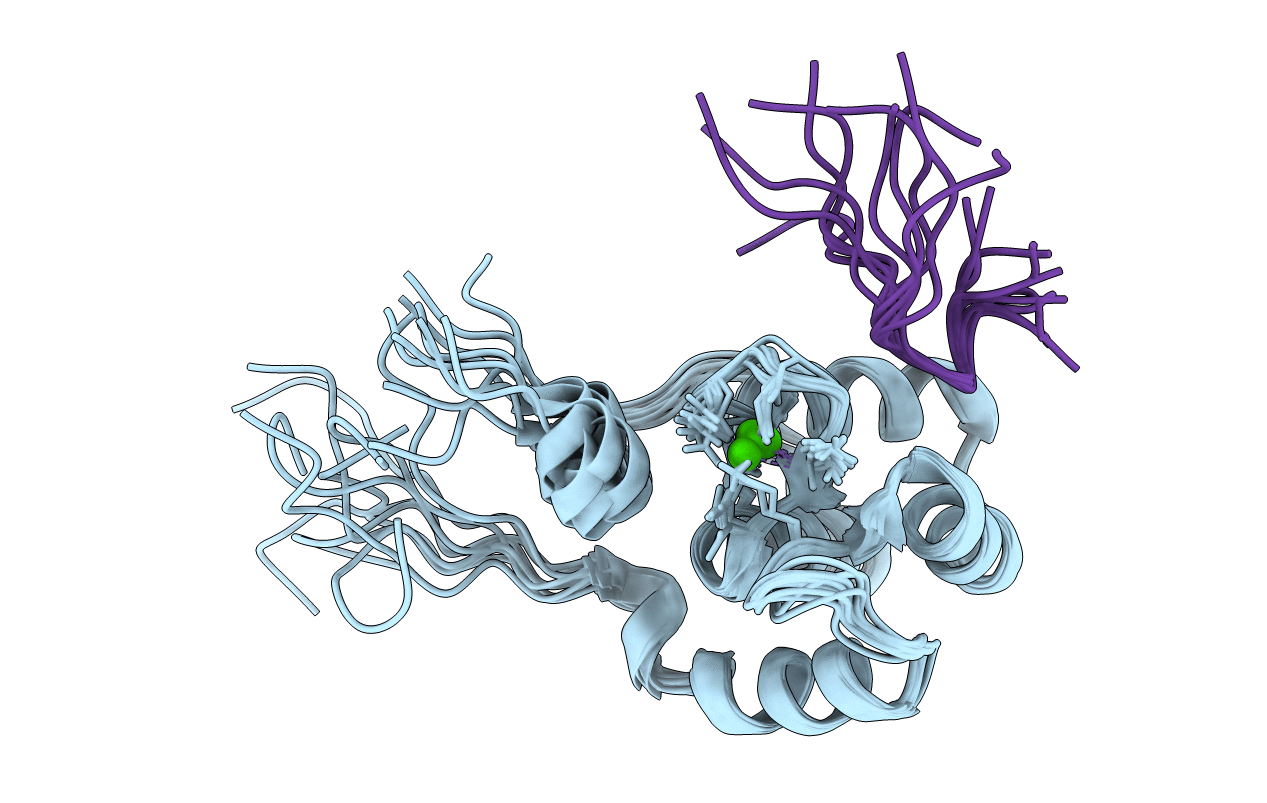
Structure of the C-terminal domain of EHD1 in complex with FNYESTDPFTAK
Biological Source:
Source Organism:
Homo sapiens (Taxon ID: 9606)
(Taxon ID: )
Host Organism:
Method Details:
Experimental Method:
Conformers Calculated:
30
Conformers Submitted:
10
Selection Criteria:
structures with the lowest energy
Macromolecular Entities
Polymer Type:polypeptide(L)
Molecule:EH domain-containing protein 1
Gene (Uniprot):EHD1
Chain IDs:A
Chain Length:105
Number of Molecules:1
Biological Source:Homo sapiens
Polymer Type:polypeptide(L)
Molecule:Rab11-FIP2 DPF peptide FNYESTDPFTAK
Chain IDs:B
Chain Length:12
Number of Molecules:1
Biological Source:
Ligand Molecules
Primary Citation
Structural insight into the interaction of proteins containing NPF, DPF, and GPF motifs with the C-terminal EH-domain of EHD1.
Protein Sci. 18 2471 2479 (2009)
PMID: 19798736 DOI: 10.1002/pro.258

Abstact

Eps15 homology (EH)-domain containing proteins are regulators of endocytic membrane trafficking. EH-domain binding to proteins containing the tripeptide NPF has been well characterized, but recent studies have shown that EH-domains are also able to interact with ligands containing DPF or GPF motifs. We demonstrate that the three motifs interact in a similar way with the EH-domain of EHD1, with the NPF motif having the highest affinity due to the presence of an intermolecular hydrogen bond. The weaker affinity for the DPF and GPF motifs suggests that if complex formation occurs in vivo, they may require high ligand concentrations, the presence of successive motifs and/or specific flanking residues.

Legend

Protein

Chemical

Disease

Primary Citation of related structures