2KDF image
Entry Detail
PDB ID:
2KDF
Keywords:
Title:
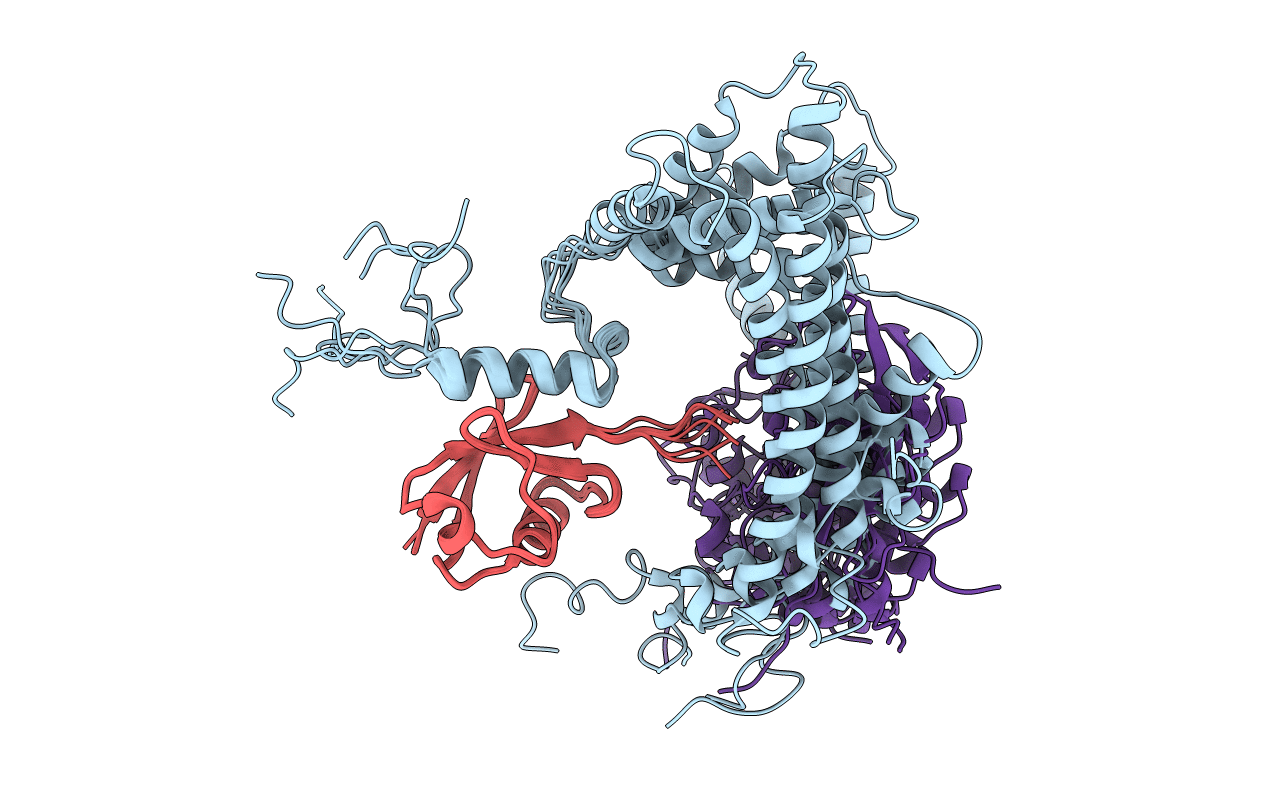
NMR structure of minor S5a (196-306):K48 linked diubiquitin species
Biological Source:
Source Organism:
Host Organism:
PDB Version:
Deposition Date:
2009-01-06
Release Date:
2009-09-01
Method Details:
Experimental Method:
Conformers Calculated:
30
Conformers Submitted:
7
Selection Criteria:
structures with the lowest energy
Macromolecular Entities
Polymer Type:polypeptide(L)
Description:26S proteasome non-ATPase regulatory subunit 4
Chain IDs:A
Chain Length:111
Number of Molecules:1
Biological Source:Homo sapiens
Polymer Type:polypeptide(L)
Description:Ubiquitin
Chain IDs:B, C
Chain Length:76
Number of Molecules:2
Biological Source:Homo sapiens
Ligand Molecules
Primary Citation
Structure of the s5a:k48-linked diubiquitin complex and its interactions with rpn13.
Mol.Cell 35 280 290 (2009)
PMID: 19683493 DOI: 10.1016/j.molcel.2009.06.010

Abstact

Degradation by the proteasome typically requires substrate ubiquitination. Two ubiquitin receptors exist in the proteasome, S5a/Rpn10 and Rpn13. Whereas Rpn13 has only one ubiquitin-binding surface, S5a binds ubiquitin with two independent ubiquitin-interacting motifs (UIMs). Here, we use nuclear magnetic resonance (NMR) and analytical ultracentrifugation to define at atomic level resolution how S5a binds K48-linked diubiquitin, in which K48 of one ubiquitin subunit (the "proximal" one) is covalently bonded to G76 of the other (the "distal" subunit). We demonstrate that S5a's UIMs bind the two subunits simultaneously with a preference for UIM2 binding to the proximal subunit while UIM1 binds to the distal one. In addition, NMR experiments reveal that Rpn13 and S5a bind K48-linked diubiquitin simultaneously with subunit specificity, and a model structure of S5a and Rpn13 bound to K48-linked polyubiquitin is provided. Altogether, our data demonstrate that S5a is highly adaptive and cooperative toward binding ubiquitin chains.

Legend

Protein

Chemical

Disease

Primary Citation of related structures