2KD9 image
Entry Detail
PDB ID:
2KD9
Keywords:
Title:
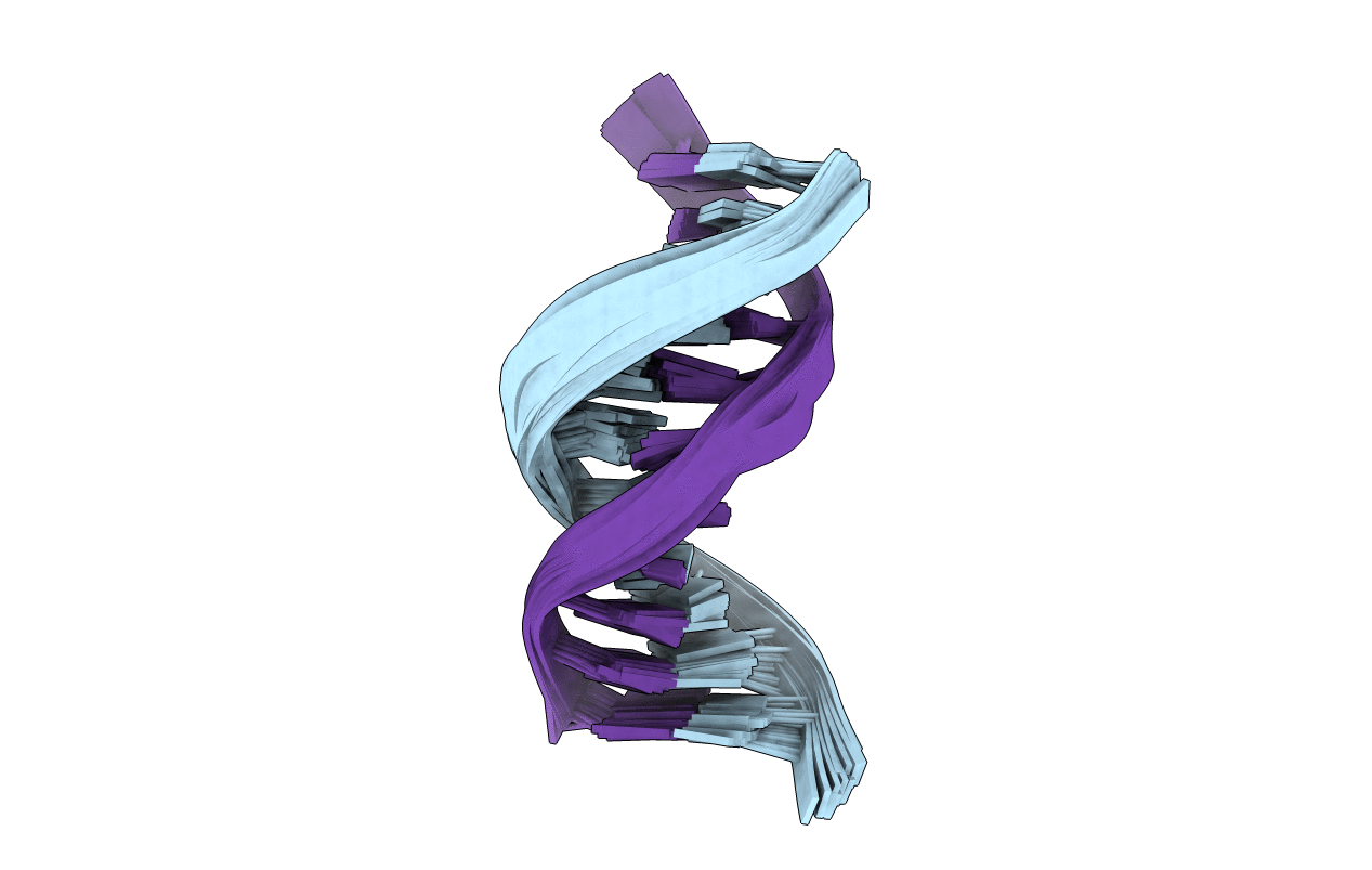
Solution Structure of DNA Containing Alpha-OH-PdG: the Mutagenic Adduct Produced by Acrolein
Biological Source:
Source Organism:
PDB Version:
Deposition Date:
2009-01-05
Release Date:
2009-04-21
Method Details:
Experimental Method:
Conformers Calculated:
25
Conformers Submitted:
26
Selection Criteria:
all calculated structures submitted
Macromolecular Entities
Polymer Type:polydeoxyribonucleotide
Description:5'-D(*CP*GP*TP*AP*CP*(63G)P*CP*AP*TP*GP*C)-3'
Chain IDs:A
Chain Length:11
Number of Molecules:1
Biological Source:
Polymer Type:polydeoxyribonucleotide
Description:5'-D(*GP*CP*AP*TP*GP*CP*GP*TP*AP*CP*G)-3'
Chain IDs:B
Chain Length:11
Number of Molecules:1
Biological Source:
Modified Residue
Compound ID Chain ID Parent Comp ID Details 2D Image
63G A DG ?
Ligand Molecules
Primary Citation
Solution structure of DNA containing alpha-OH-PdG: the mutagenic adduct produced by acrolein.
Nucleic Acids Res. 37 2153 2163 (2009)
PMID: 19223332 DOI: 10.1093/nar/gkp076

Abstact

Acrolein is a cell metabolic product and a main component of cigarette smoke. Its reaction with DNA produces two guanine lesions gamma-OH-PdG, a major adduct that is nonmutagenic in mammalian cells, and the positional isomer alpha-OH-PdG. We describe here the solution structure of a short DNA duplex containing a single alpha-OH-PdG lesion, as determined by solution NMR spectroscopy and restrained molecular dynamics simulations. The spectroscopic data show a mostly regular right-handed helix, locally perturbed at its center by the presence of the lesion. All undamaged residues of the duplex are in anti orientation, forming standard Watson-Crick base-pair alignments. Duplication of proton signals near the damaged site differentiates two enantiomeric duplexes, thus establishing the exocyclic nature of the lesion. At the lesion site, alpha-OH-PdG rotates to a syn conformation, pairing to its counter cytosine residue that is protonated at pH 5.9. Three-dimensional models produced by restrained molecular dynamics simulations show different hydrogen-bonding patterns between the lesion and its cytosine partner and identify further stabilization of alpha-OH-PdG in a syn conformation by intra-residue hydrogen bonds. We compare the alpha-OH-PdG.dC duplex structure with that of duplexes containing the analogous lesion propano-dG and discuss the implications of our findings for the mutagenic bypass of acrolein lesions.

Legend

Protein

Chemical

Disease

Primary Citation of related structures