2KCF image
Deposition Date 2008-12-19
Release Date 2009-01-13
Last Version Date 2024-05-22
Entry Detail
PDB ID:
2KCF
Keywords:
Title:
The NMR solution structure of the isolated Apo Pin1 WW domain
Biological Source:
Source Organism:
Homo sapiens (Taxon ID: 9606)
Host Organism:
Method Details:
Experimental Method:
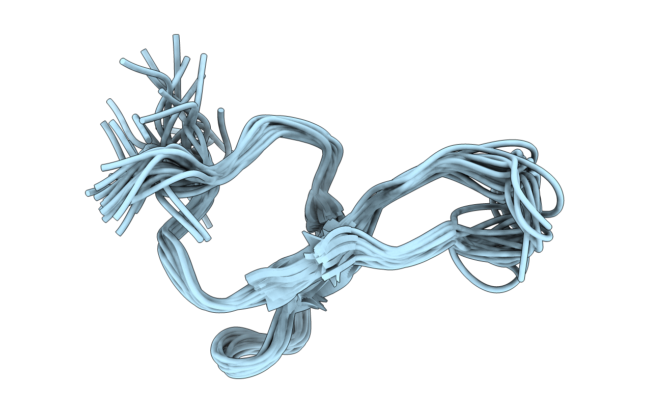
Conformers Calculated:
20
Conformers Submitted:
20
Selection Criteria:
structures with the lowest energy
Macromolecular Entities
Structures with similar UniProt ID
Protein Blast
Polymer Type:polypeptide(L)
Molecule:Peptidyl-prolyl cis-trans isomerase NIMA-interacting 1
Gene (Uniprot):PIN1
Chain IDs:A
Chain Length:36
Number of Molecules:1
Biological Source:Homo sapiens
Ligand Molecules
Primary Citation
NMR solution structure of the isolated Apo Pin1 WW domain: comparison to the x-ray crystal structures of Pin1
Biopolymers 63 111 121 (2002)
PMID: 11786999

Abstact

The NMR solution structure of the isolated Apo Pin1 WW domain (6-39) reveals that it adopts a twisted three-stranded antiparallel beta-sheet conformation, very similar to the structure exhibited by the crystal of this domain in the context of the two domain Pin1 protein. While the B factors in the apo x-ray crystal structure indicate that loop 1 and loop 2 are conformationally well defined, the solution NMR data suggest that loop 1 is quite flexible, at least in the absence of the ligand. The NMR chemical shift and nuclear Overhauser effect pattern exhibited by the 6-39 Pin1 WW domain has proven to be diagnostic for demonstrating that single site variants of this domain adopt a normally folded structure. Knowledge of this type is critical before embarking on time-consuming kinetic and thermodynamic studies required for a detailed understanding of beta-sheet folding.

Legend

Protein

Chemical

Disease

Primary Citation of related structures