
Deposition Date
2008-11-21
Release Date
2009-06-16
Last Version Date
2024-05-22
Entry Detail
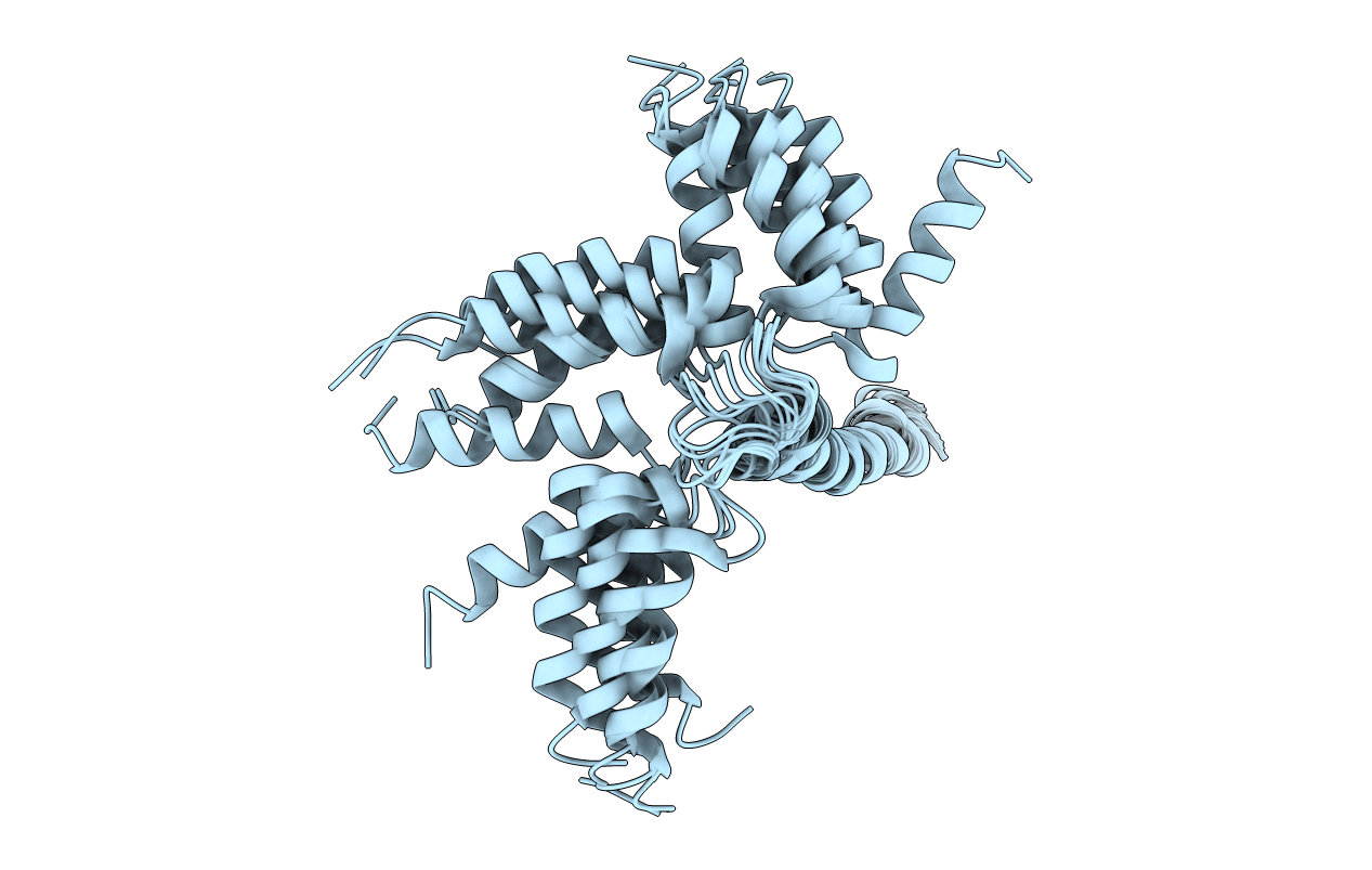
PDB ID:
2KB7
Keywords:
Title:
Hybrid solution and solid-state NMR structure of monomeric phospholamban in lipid bilayers
Biological Source:
Source Organism(s):
Escherichia coli (Taxon ID: 562)
Expression System(s):
Method Details:
Experimental Method:
Conformers Calculated:
200
Conformers Submitted:
20
Selection Criteria:
structures with the least restraint violations
