2KAU image
Deposition Date 1995-02-16
Release Date 1995-07-10
Last Version Date 2024-06-05
Entry Detail
PDB ID:
2KAU
Keywords:
Title:
THE CRYSTAL STRUCTURE OF UREASE FROM KLEBSIELLA AEROGENES AT 2.2 ANGSTROMS RESOLUTION
Biological Source:
Source Organism:
Host Organism:
Method Details:
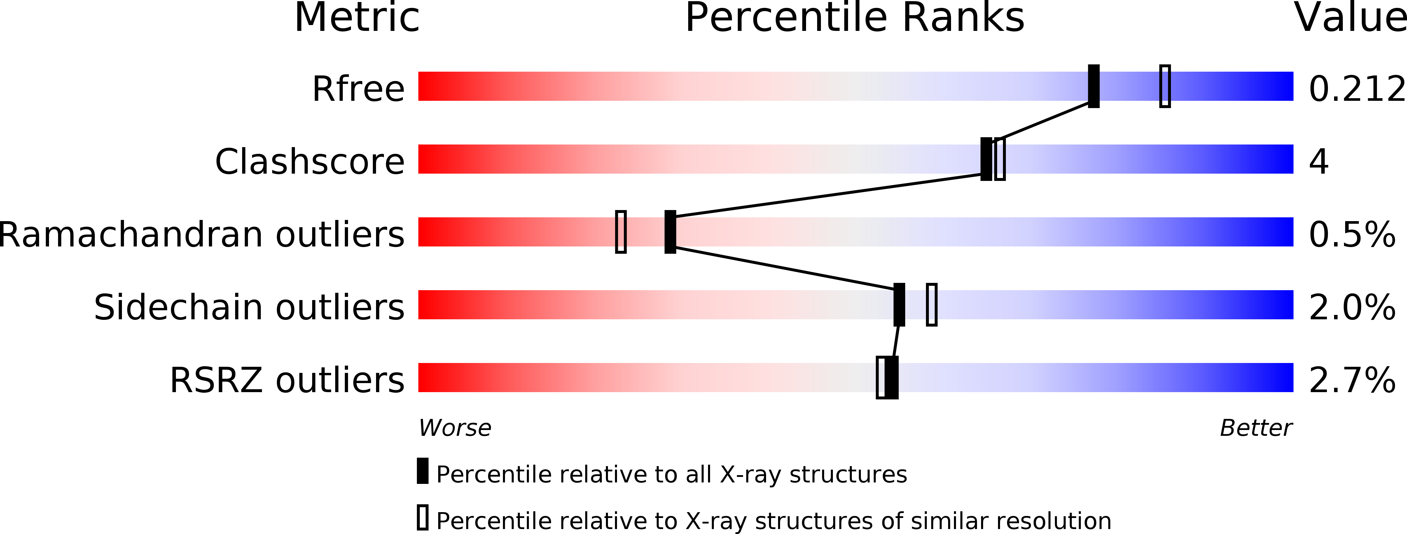
Experimental Method:
Resolution:
2.00 Å
R-Value Free:
0.22
R-Value Work:
0.18
R-Value Observed:
0.18
Space Group:
I 21 3
Macromolecular Entities
Polymer Type:polypeptide(L)
Molecule:UREASE (GAMMA CHAIN)
Gene (Uniprot):ureA
Chain IDs:A
Chain Length:100
Number of Molecules:1
Biological Source:Klebsiella aerogenes
Polymer Type:polypeptide(L)
Molecule:UREASE (BETA CHAIN)
Gene (Uniprot):ureB
Chain IDs:B
Chain Length:106
Number of Molecules:1
Biological Source:Klebsiella aerogenes
Polymer Type:polypeptide(L)
Molecule:UREASE (ALPHA CHAIN)
Gene (Uniprot):ureC
Chain IDs:C
Chain Length:567
Number of Molecules:1
Biological Source:Klebsiella aerogenes
Modified Residue
Compound ID Chain ID Parent Comp ID Details 2D Image
KCX C LYS LYSINE NZ-CARBOXYLIC ACID
Ligand Molecules
Primary Citation
The crystal structure of urease from Klebsiella aerogenes.
Science 268 998 1004 (1995)
PMID: 7754395

Abstact

The crystal structure of urease from Klebsiella aerogenes has been determined at 2.2 A resolution and refined to an R factor of 18.2 percent. The enzyme contains four structural domains: three with novel folds playing structural roles, and an (alpha beta)8 barrel domain, which contains the bi-nickel center. The two active site nickels are 3.5 A apart. One nickel ion is coordinated by three ligands (with low occupancy of a fourth ligand) and the second is coordinated by five ligands. A carbamylated lysine provides an oxygen ligand to each nickel, explaining why carbon dioxide is required for the activation of urease apoenzyme. The structure is compatible with a catalytic mechanism whereby urea ligates Ni-1 to complete its tetrahedral coordination and a hydroxide ligand of Ni-2 attacks the carbonyl carbon. A surprisingly high structural similarity between the urease catalytic domain and that of the zinc-dependent adenosine deaminase reveals a remarkable example of active site divergence.

Legend

Protein

Chemical

Disease

Primary Citation of related structures