Deposition Date
2008-08-28
Release Date
2009-11-10
Last Version Date
2024-05-22
Entry Detail
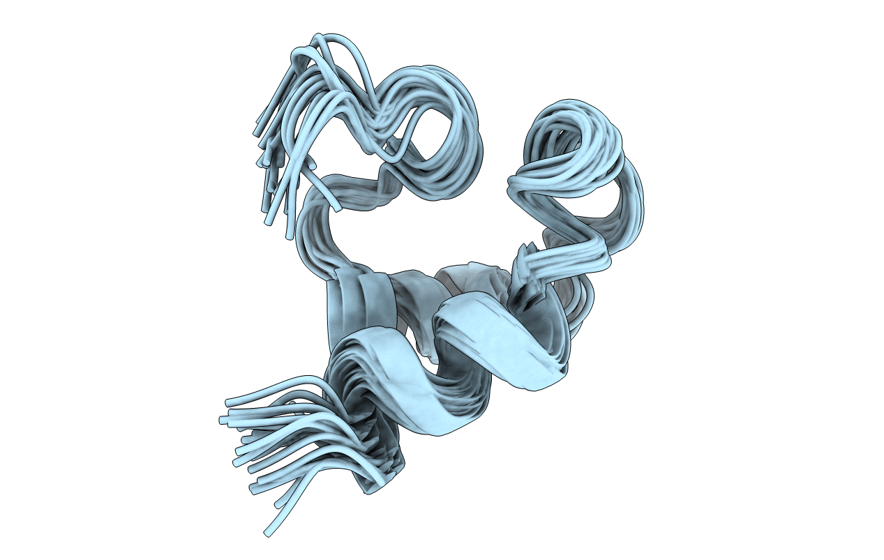
PDB ID:
2K7Y
Keywords:
Title:
Solution fold of HIV-1 Virus protein U cytoplasmic domain in the presence of DPC micelles
Biological Source:
Source Organism(s):
Human immunodeficiency virus type 1 (Taxon ID: 11691)
Expression System(s):
Method Details:
Experimental Method:
Conformers Calculated:
100
Conformers Submitted:
20
Selection Criteria:
target function