Deposition Date
2008-06-25
Release Date
2008-07-08
Last Version Date
2024-05-29
Entry Detail
PDB ID:
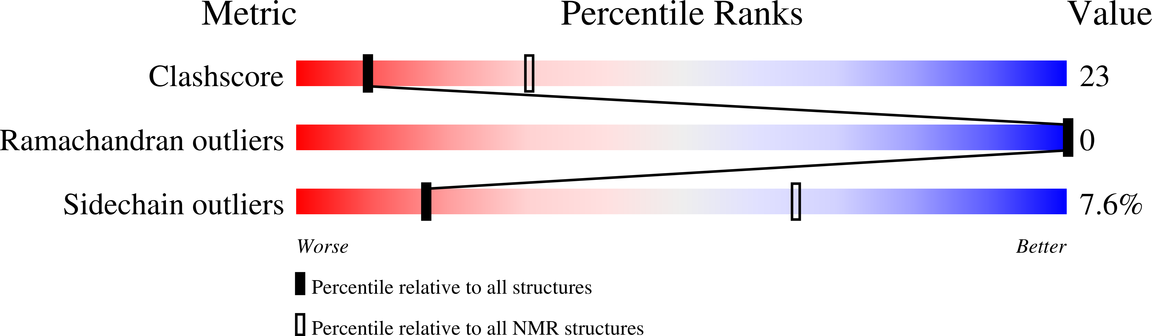
2K59
Keywords:
Title:
NMR structures of the second transmembrane domain of the neuronal acetylcholine receptor beta 2 subunit
Method Details:
Experimental Method:
Conformers Calculated:
100
Conformers Submitted:
20
Selection Criteria:
structures with the lowest energy