2K4R image
Deposition Date 2008-06-16
Release Date 2008-12-09
Last Version Date 2024-11-27
Entry Detail
PDB ID:
2K4R
Keywords:
Title:
NMR solution structure of the neurotrypsin kringle domain
Biological Source:
Source Organism(s):
Expression System(s):
Method Details:
Experimental Method:
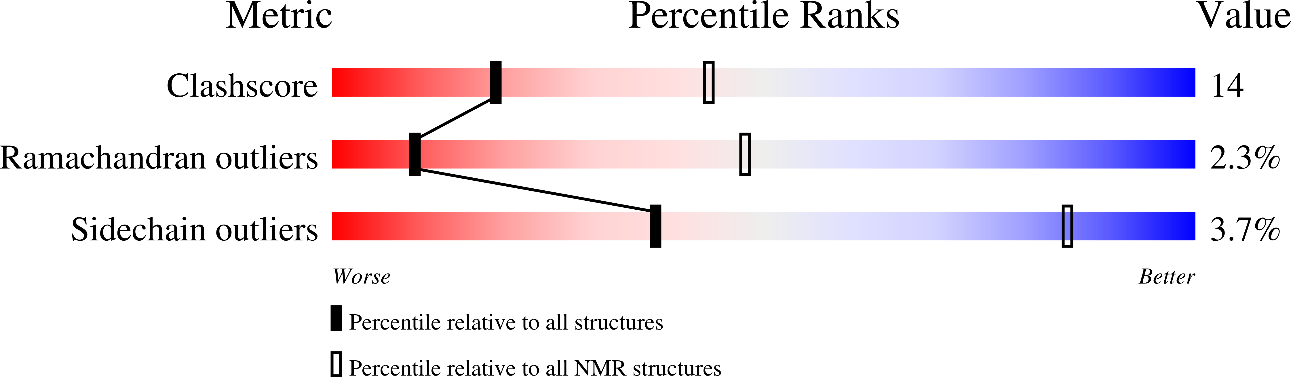
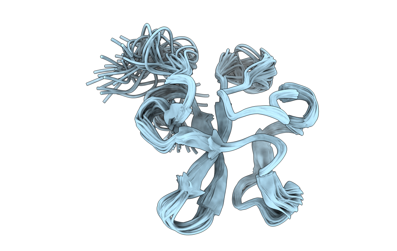
Conformers Calculated:
70
Conformers Submitted:
29
Selection Criteria:
STRUCTURES WITH THE LEAST RESTRAINT VIOLATIONS, STRUCTURES WITH THE LOWEST ENERGY
Macromolecular Entities
Structures with similar UniProt ID
Protein Blast
Polymer Type:polypeptide(L)
Molecule:Neurotrypsin
Chain IDs:A
Chain Length:77
Number of Molecules:1
Biological Source:Rattus norvegicus
Ligand Molecules
Primary Citation
NMR Solution Structure of the Neurotrypsin Kringle Domain
Biochemistry 47 12290 12298 (2008)
PMID: 18956887 DOI: 10.1021/bi800555z

Abstact

Neurotrypsin is a multidomain protein that serves as a brain-specific serine protease. Here we report the NMR structure of its kringle domain, NT/K. The data analysis was performed with the BACUS (Bayesian analysis of coupled unassigned spins) algorithm. This study presents the first application of BACUS to the structure determination of a 13C unenriched protein for which no prior experimental 3D structure was available. NT/K adopts the kringle fold, consisting of an antiparallel beta-sheet bridged by an overlapping pair of disulfides. The structure reveals the presence of a surface-exposed left-handed polyproline II helix that is closely packed to the core beta-structure. This feature distinguishes NT/K from other members of the kringle fold and points toward a novel functional role for a kringle domain. Functional divergence among kringle domains is discussed on the basis of their surface and electrostatic characteristics.

Legend

Protein

Chemical

Disease

Primary Citation of related structures
Feedback Form
Name
Email
Institute
Feedback