2K2O image
Deposition Date 2008-04-07
Release Date 2008-06-10
Last Version Date 2024-05-01
Entry Detail
PDB ID:
2K2O
Title:
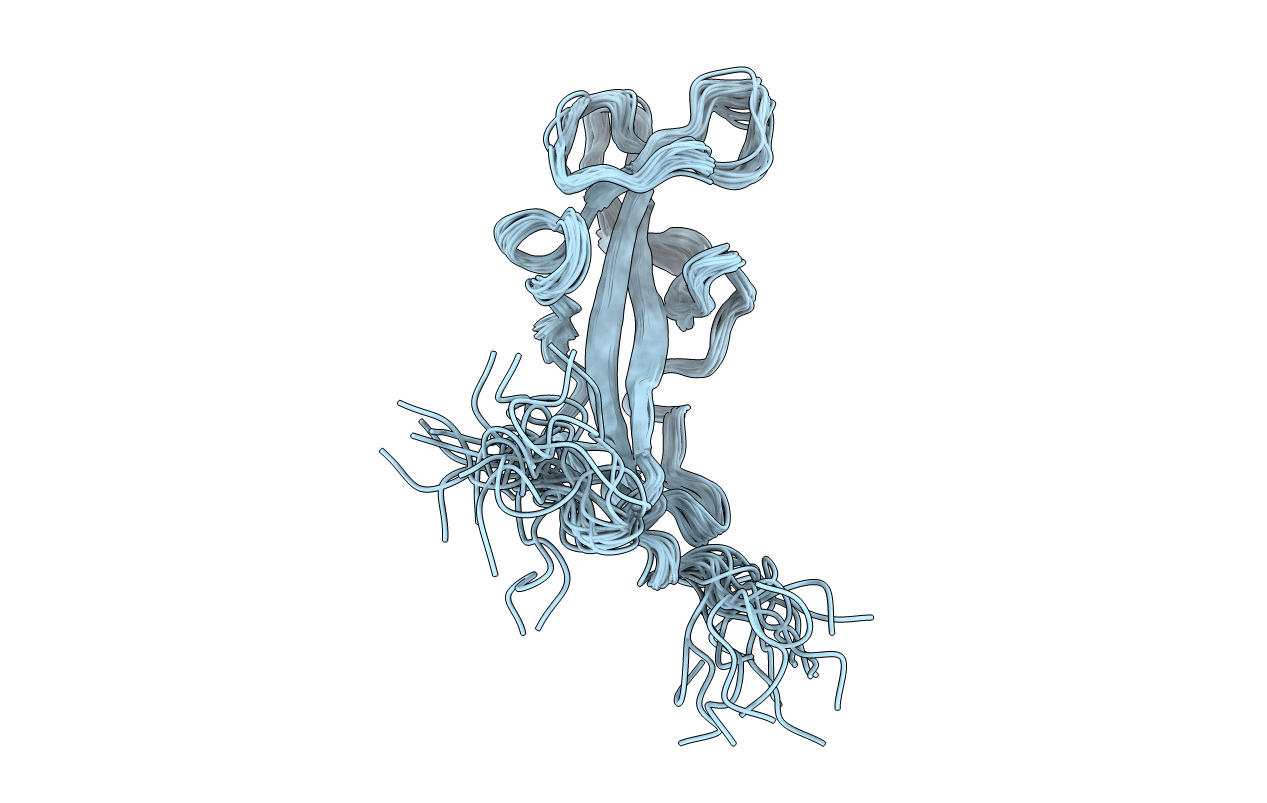
Solution Structure of the inner DysF domain of human myoferlin
Biological Source:
Source Organism:
Homo sapiens (Taxon ID: )
Host Organism:
Method Details:
Experimental Method:
Conformers Calculated:
100
Conformers Submitted:
20
Selection Criteria:
structures with the lowest energy
Macromolecular Entities
Structures with similar UniProt ID
Protein Blast
Polymer Type:polypeptide(L)
Molecule:Myoferlin
Gene (Uniprot):MYOF
Chain IDs:A
Chain Length:123
Number of Molecules:1
Biological Source:Homo sapiens
Ligand Molecules
Primary Citation
Solution structure of the inner DysF domain of myoferlin and implications for limb girdle muscular dystrophy type 2b.
J.Mol.Biol. 379 981 990 (2008)
PMID: 18495154 DOI: 10.1016/j.jmb.2008.04.046

Abstact

Mutations in the protein dysferlin, a member of the ferlin family, lead to limb girdle muscular dystrophy type 2B and Myoshi myopathy. The ferlins are large proteins characterised by multiple C2 domains and a single C-terminal membrane-spanning helix. However, there is sequence conservation in some of the ferlin family in regions outside the C2 domains. In one annotation of the domain structure of these proteins, an unusual internal duplication event has been noted where a putative domain is inserted in between the N- and C-terminal parts of a homologous domain. This domain is known as the DysF domain. Here, we present the solution structure of the inner DysF domain of the dysferlin paralogue myoferlin, which has a unique fold held together by stacking of arginine and tryptophans, mutations that lead to clinical disease in dysferlin.

Legend

Protein

Chemical

Disease

Primary Citation of related structures
Feedback Form
Name
Email
Institute
Feedback