2K0G image
Deposition Date 2008-02-02
Release Date 2009-02-10
Last Version Date 2024-05-29
Entry Detail
PDB ID:
2K0G
Title:
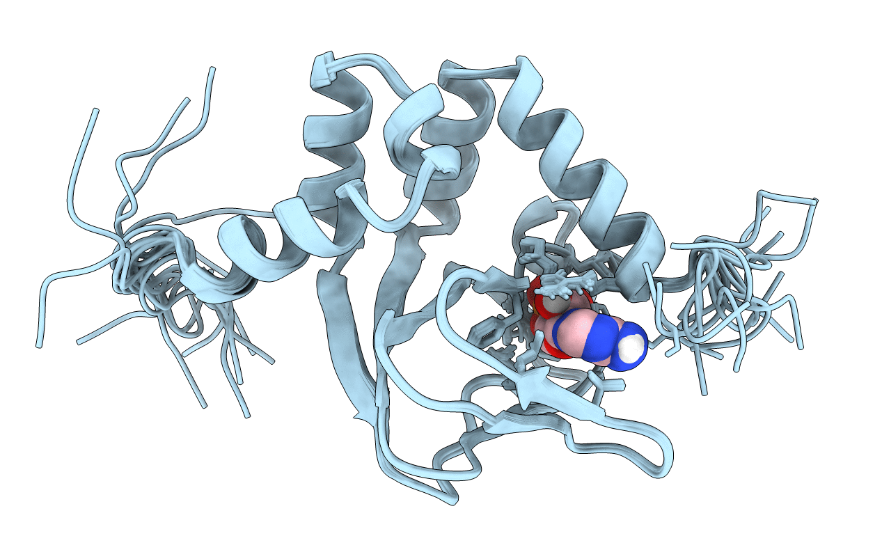
Solution Structure of a Bacterial Cyclic Nucleotide-Activated K+ Channel Binding Domain in Complex with cAMP
Biological Source:
Source Organism:
Rhizobium loti (Taxon ID: )
Host Organism:
Method Details:
Experimental Method:
Conformers Calculated:
100
Conformers Submitted:
15
Selection Criteria:
15 structures with the lowest energy
Macromolecular Entities
Structures with similar UniProt ID
Protein Blast
Polymer Type:polypeptide(L)
Molecule:Mll3241 protein
Gene (Uniprot):mll3241
Chain IDs:A
Chain Length:142
Number of Molecules:1
Biological Source:Rhizobium loti
Ligand Molecules
Primary Citation
Solution structure of the Mesorhizobium loti K1 channel cyclic nucleotide-binding domain in complex with cAMP.
Embo Rep. 10 729 735 (2009)
PMID: 19465888 DOI: 10.1038/embor.2009.68

Abstact

Cyclic nucleotide-sensitive ion channels, known as HCN and CNG channels, are crucial in neuronal excitability and signal transduction of sensory cells. HCN and CNG channels are activated by binding of cyclic nucleotides to their intracellular cyclic nucleotide-binding domain (CNBD). However, the mechanism by which the binding of cyclic nucleotides opens these channels is not well understood. Here, we report the solution structure of the isolated CNBD of a cyclic nucleotide-sensitive K(+) channel from Mesorhizobium loti. The protein consists of a wide anti-parallel beta-roll topped by a helical bundle comprising five alpha-helices and a short 3(10)-helix. In contrast to the dimeric arrangement ('dimer-of-dimers') in the crystal structure, the solution structure clearly shows a monomeric fold. The monomeric structure of the CNBD supports the hypothesis that the CNBDs transmit the binding signal to the channel pore independently of each other.

Legend

Protein

Chemical

Disease

Primary Citation of related structures