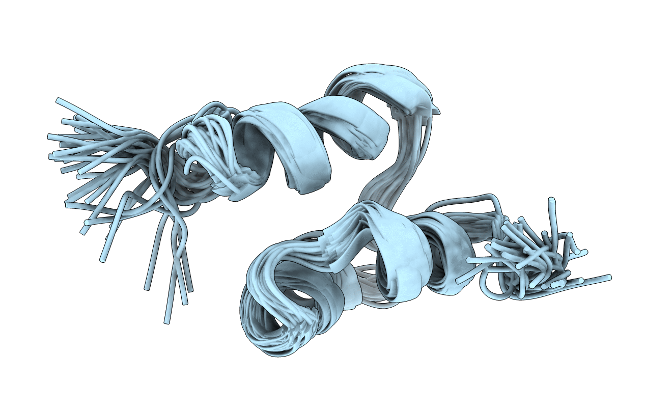
2K0B image
Deposition Date 2008-01-31
Release Date 2008-02-19
Last Version Date 2024-05-29
Entry Detail
PDB ID:
2K0B
Title:
NMR structure of the UBA domain of p62 (SQSTM1)
Biological Source:
Source Organism:
Homo sapiens (Taxon ID: 9606)
Method Details:
Experimental Method:
Conformers Calculated:
100
Conformers Submitted:
30
Selection Criteria:
structures with the lowest energy
Macromolecular Entities
Structures with similar UniProt ID
Protein Blast
Polymer Type:polypeptide(L)
Molecule:Sequestosome-1
Gene (Uniprot):SQSTM1
Chain IDs:A (auth: X)
Chain Length:52
Number of Molecules:1
Biological Source:Homo sapiens
Ligand Molecules
Primary Citation
Conformation and dynamics of the three-helix bundle UBA domain of p62 from experiment and simulation.
Proteins 71 227 240 (2007)
PMID: 17932931 DOI: 10.1002/prot.21692

Abstact

The ubiquitin associated domain of p62 is a small three-helix bundle of approximately 50 residues that mediates the recognition of polyubiquitin chains and ubiquitylated substrates. The solution structure of a 52 residue construct containing this domain has been characterized using heteronuclear nuclear magnetic resonance (NMR) methods. The resulting ensemble of NMR-derived structures was used in molecular dynamics (MD) simulations to investigate the equilibrium conformation and dynamics of this domain. NOE and (15)N relaxation data have been used to validate the structural ensemble produced by the MD simulations and show a good correlation for residues in regions of secondary structure. A similar approach was taken using an ensemble of structures from the MD simulations to calculate electronic circular dichroism (CD) and IR spectra from first principles with an encouraging correlation with the experimental CD and IR data.

Legend

Protein

Chemical

Disease

Primary Citation of related structures