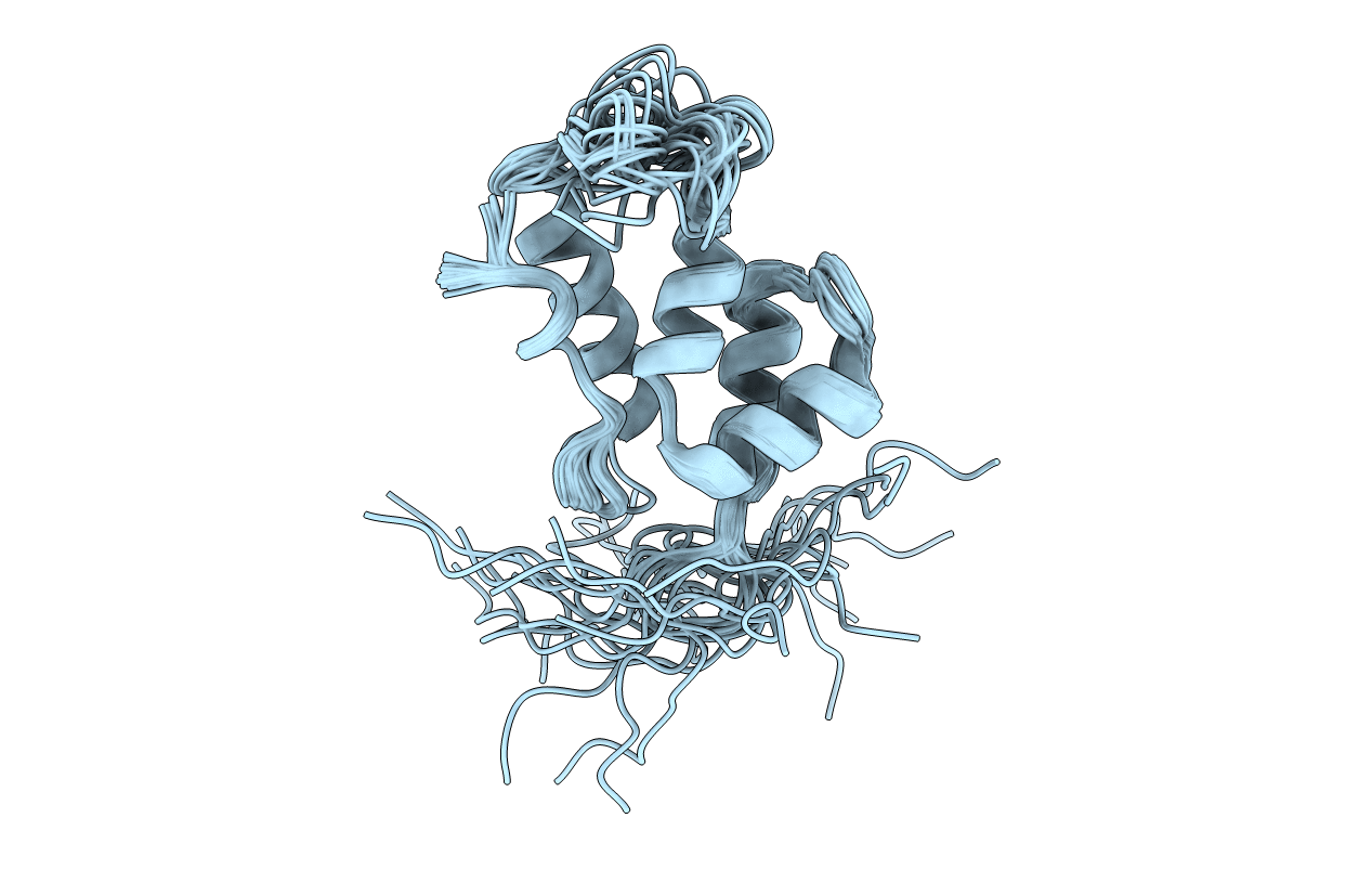
2JYG image
Deposition Date 2007-12-13
Release Date 2008-02-26
Last Version Date 2024-05-29
Entry Detail
PDB ID:
2JYG
Keywords:
Title:
Solution Structure of the W184A/M185A Mutant of the Carboxy-terminal Dimerization Domain of the HIV-1 Capsid Protein
Biological Source:
Source Organism:
Host Organism:
Method Details:
Experimental Method:
Conformers Calculated:
500
Conformers Submitted:
30
Selection Criteria:
target function
Macromolecular Entities
Polymer Type:polypeptide(L)
Molecule:Capsid protein p24 (CA)
Gene (Uniprot):gag-pol
Chain IDs:A
Chain Length:84
Number of Molecules:1
Biological Source:Human immunodeficiency virus 1
Ligand Molecules
Primary Citation
Solution Structure of a Double Mutant of the Carboxy-Terminal Dimerization Domain of the HIV-1 Capsid Protein.
Biochemistry 47 2289 2297 (2008)
PMID: 18220423 DOI: 10.1021/bi7022128

Abstact

As in other retroviruses, the HIV-1 capsid (CA) protein is composed of two domains, the N-terminal domain (NTD) and the C-terminal domain (CTD), joined by a flexible linker. The dimerization of the CTD is thought to be a critical step in the assembly of the immature and mature viral capsids. The precise nature of the functional form of CTD dimerization interface has been a subject of considerable interest. Previously, the CTD dimer was thought to involve a face-to-face dimerization observed in the early crystallographic studies. Recently, the crystallographic structure for a domain-swapped CTD dimer has been determined. This dimer, with an entirely different interface that includes the major homology region (MHR) has been suggested as the functional form during the Gag assembly. The structure determination of the monomeric wt CTD of HIV-1 has not been possible because of the monomer-dimer equilibrium in solution. We report the NMR structure of the [W184A/M185A]-CTD mutant in its monomeric form. These mutations interfere with dimerization without abrogating the assembly activity of Gag and CA. The NMR structure shows some important differences compared to the CTD structure in the face-to-face dimer. Notably, the helix-2 is much shorter, and the kink seen in the crystal structure of the wt CTD in the face-to-face dimer is absent. These NMR studies suggest that dimerization-induced conformational changes may be present in the two crystal structures of the CTD dimers and also suggest a mechanism that can simultaneously accommodate both of the distinctly different dimer models playing functional roles during the Gag assembly of the immature capsids.

Legend

Protein

Chemical

Disease

Primary Citation of related structures