
Deposition Date
2007-12-07
Release Date
2007-12-18
Last Version Date
2024-05-01
Entry Detail
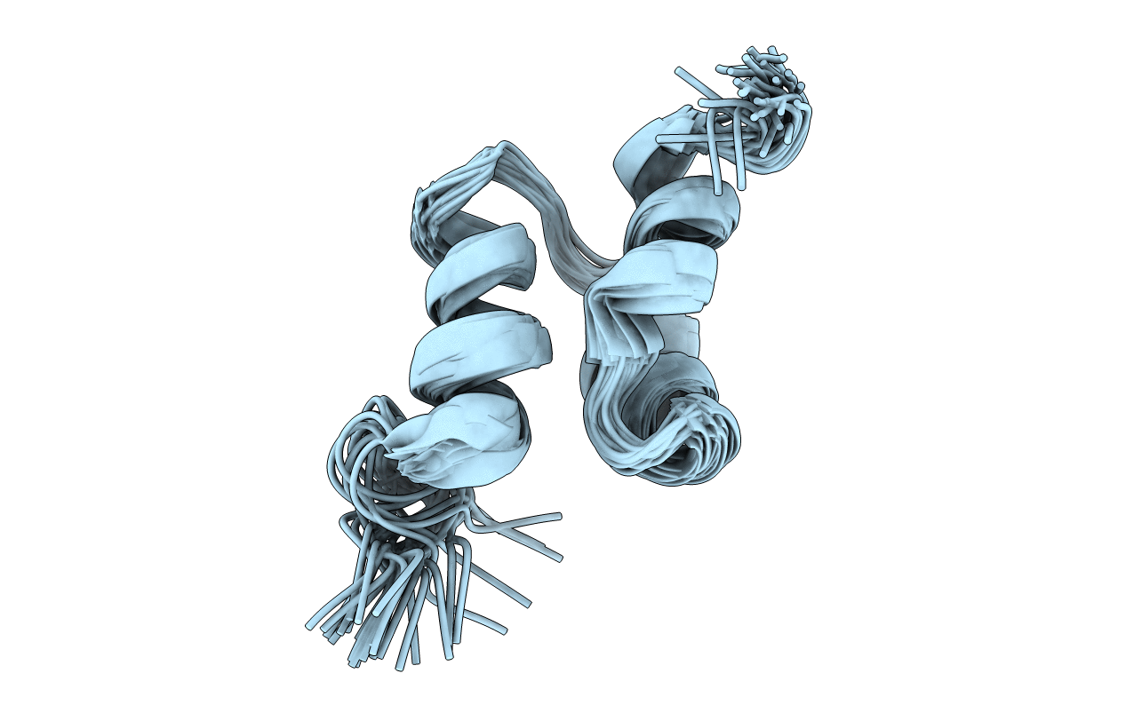
PDB ID:
2JY7
Keywords:
Title:
NMR structure of the ubiquitin associated (UBA) domain of p62 (SQSTM1). RDC refined
Biological Source:
Source Organism(s):
Homo sapiens (Taxon ID: 9606)
Expression System(s):
Method Details:
Experimental Method:
Conformers Calculated:
100
Conformers Submitted:
30
Selection Criteria:
structures with the lowest energy
