Deposition Date
2007-11-30
Release Date
2008-03-11
Last Version Date
2024-05-15
Entry Detail
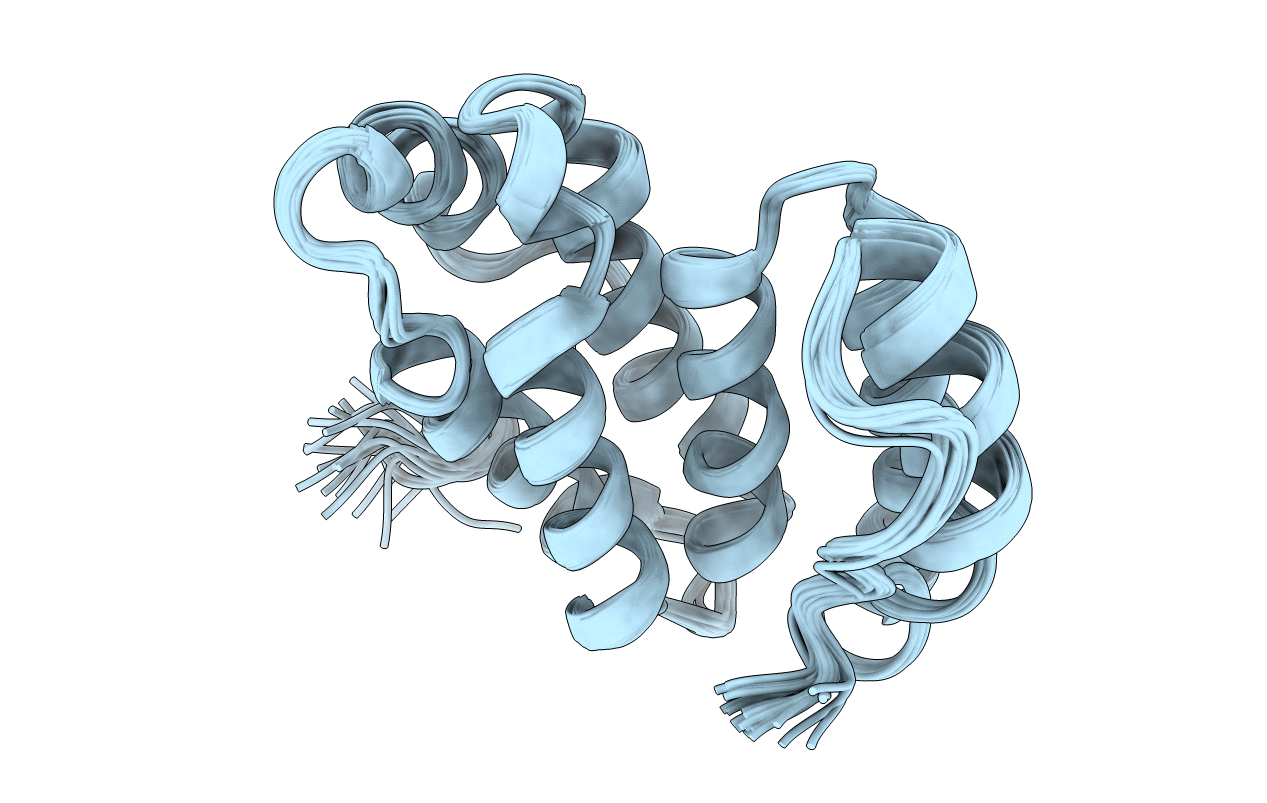
PDB ID:
2JXU
Keywords:
Title:
NMR solution structure of KP-TerB, a tellurite resistance protein from Klebsiella pneumoniae
Biological Source:
Source Organism(s):
Klebsiella pneumoniae (Taxon ID: 484021)
Expression System(s):
Method Details:
Experimental Method:
Conformers Calculated:
200
Conformers Submitted:
20
Selection Criteria:
structures with the least restraint violations