2JXH image
Deposition Date 2007-11-19
Release Date 2008-10-21
Last Version Date 2024-05-29
Entry Detail
PDB ID:
2JXH
Title:
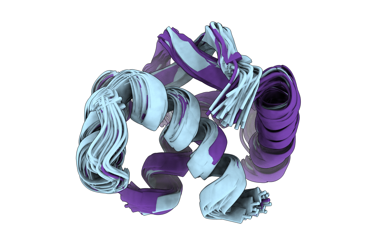
Solution Structure of DNA binding domain of Proline Utilization A (PutA) for Psuedomonas putida
Biological Source:
Source Organism:
Method Details:
Experimental Method:
Conformers Calculated:
100
Conformers Submitted:
30
Selection Criteria:
structures with the lowest energy
Macromolecular Entities
Structures with similar UniProt ID
Protein Blast
Polymer Type:polypeptide(L)
Molecule:Proline dehydrogenase
Gene (Uniprot):putA
Chain IDs:A, B
Chain Length:45
Number of Molecules:2
Biological Source:Pseudomonas putida
Ligand Molecules
Primary Citation
Solution structure of the Pseudomonas putida protein PpPutA45 and its DNA complex
Proteins 75 12 27 (2008)
PMID: 18767154 DOI: 10.1002/prot.22217

Abstact

Proline utilization A (PutA) is a membrane-associated multifunctional enzyme that catalyzes the oxidation of proline to glutamate in a two-step process. In certain, gram-negative bacteria such as Pseudomonas putida, PutA also acts as an auto repressor in the cytoplasm, when an insufficient concentration of proline is available. Here, the N-terminal residues 1-45 of PutA from P. putida (PpPutA45) are shown to be responsible for DNA binding and dimerization. The solution structure of PpPutA45 was determined using NMR methods, where the protein is shown to be a symmetrical homodimer (12 kDa) consisting of two ribbon-helix-helix (RHH) structures. DNA sequence recognition by PpPutA45 was determined using DNA gel mobility shift assays and NMR chemical shift perturbations (CSPs). PpPutA45 was shown to bind a 14 base-pair DNA oligomer (5'-GCGGTTGCACCTTT-3'). A model of the PpPutA45-DNA oligomer complex was generated using Haddock 2.1. The antiparallel beta-sheet that results from PpPutA45 dimerization serves as the DNA recognition binding site by inserting into the DNA major groove. The dimeric core of four alpha-helices provides a structural scaffold for the beta-sheet from which residues Thr5, Gly7, and Lys9 make sequence-specific contacts with the DNA. The structural model implies flexibility of Lys9 which can make hydrogen bond contacts with either guanine or thymine. The high sequence and structure conservation of the PutA RHH domain suggest interdomain interactions play an important role in the evolution of the protein.

Legend

Protein

Chemical

Disease

Primary Citation of related structures