2JWN image
Deposition Date 2007-10-16
Release Date 2007-10-30
Last Version Date 2024-05-29
Entry Detail
PDB ID:
2JWN
Title:
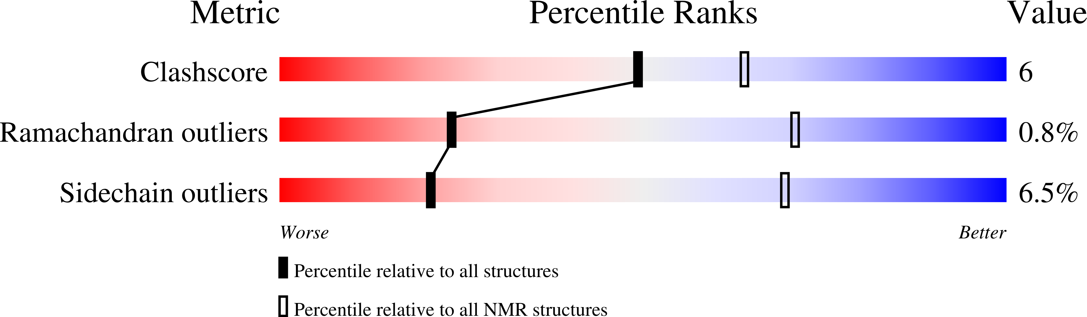
Solution NMR structure of the protease-resistent domain of Xenopus laevis ePABP2
Biological Source:
Source Organism:
Xenopus laevis (Taxon ID: 8355)
Host Organism:
Method Details:
Experimental Method:
Conformers Calculated:
100
Conformers Submitted:
20
Selection Criteria:
target function
Macromolecular Entities
Structures with similar UniProt ID
Protein Blast
Polymer Type:polypeptide(L)
Molecule:Embryonic polyadenylate-binding protein 2-B
Gene (Uniprot):Pabpn1l-b
Chain IDs:A, B
Chain Length:124
Number of Molecules:2
Biological Source:Xenopus laevis
Ligand Molecules
Primary Citation
Structural basis for RNA recognition by a type II poly(A)-binding protein.
Proc.Natl.Acad.Sci.USA 105 15317 15322 (2008)
PMID: 18824697 DOI: 10.1073/pnas.0801274105

Abstact

We identified a functional domain (XlePABP2-TRP) of Xenopus laevis embryonic type II poly(A)-binding protein (XlePABP2). The NMR structure of XlePABP2-TRP revealed that the protein is a homodimer formed by the antiparallel association of beta-strands from the single RNA recognition motif (RRM) domain of each subunit. In each subunit of the homodimer, the canonical RNA recognition site is occluded by a polyproline motif. Upon poly(A) binding, XlePABP2-TRP undergoes a dimer-monomer transition that removes the polyproline motif from the RNA recognition site and allows it to be replaced by the adenosine nucleotides of poly(A). Our results provide high-resolution structural information concerning type II PABPs and an example of a single RRM domain protein that transitions from a homodimer to a monomer upon RNA binding. These findings advance our understanding of RRM domain regulation, poly(A) recognition, and are relevant to understanding how type II PABPs function in mRNA processing and human disease.

Legend

Protein

Chemical

Disease

Primary Citation of related structures
Feedback Form
Name
Email
Institute
Feedback