
Deposition Date
2007-10-15
Release Date
2008-04-01
Last Version Date
2024-05-29
Entry Detail
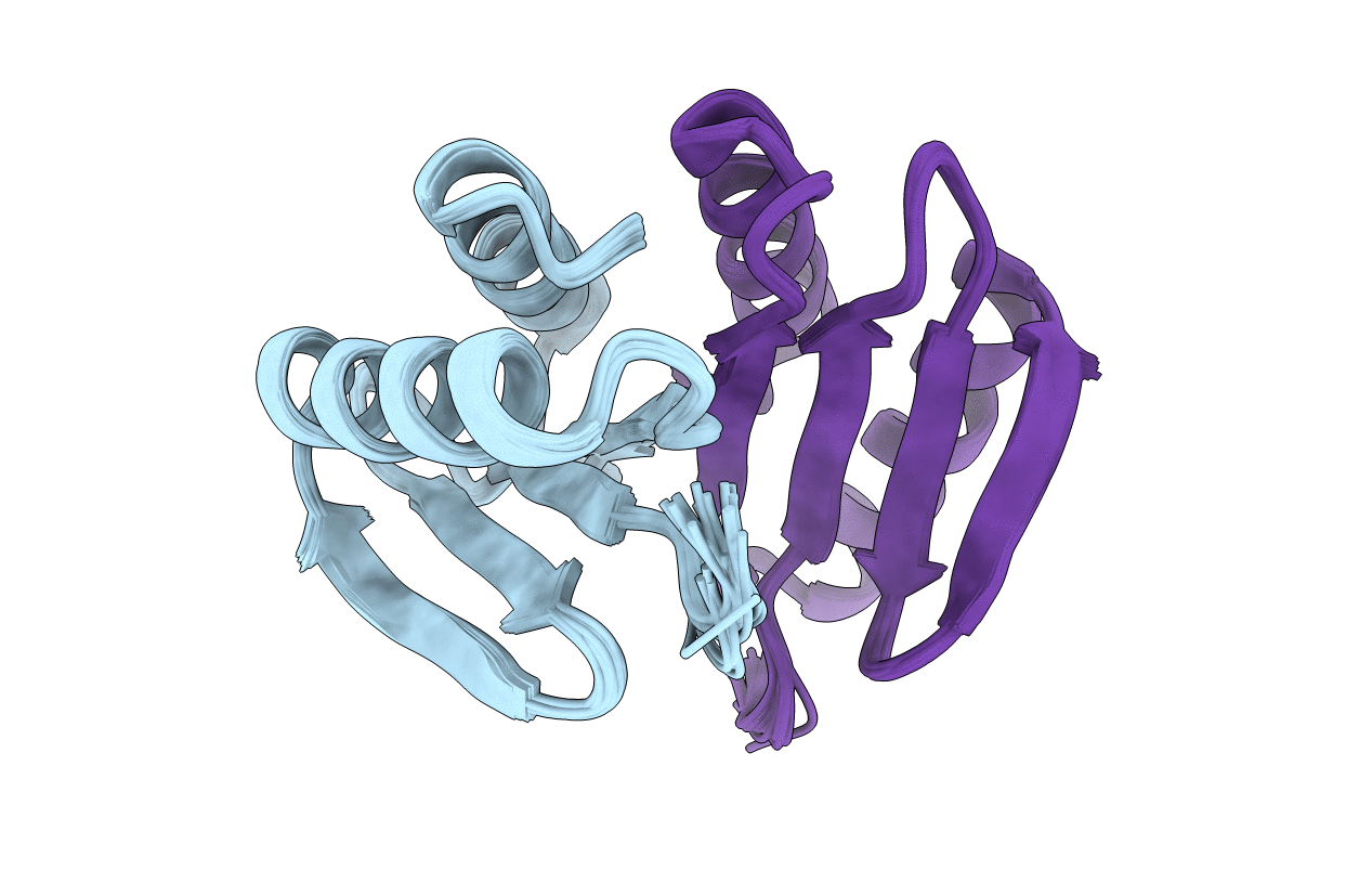
PDB ID:
2JWK
Keywords:
Title:
Solution Structure of the periplasmic domain of TolR from Haemophilus influenzae
Biological Source:
Source Organism(s):
Haemophilus influenzae (Taxon ID: 727)
Expression System(s):
Method Details:
Experimental Method:
Conformers Calculated:
40
Conformers Submitted:
20
Selection Criteria:
structures with the least restraint violations
