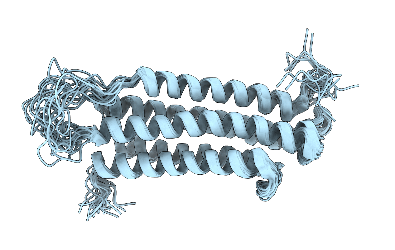
2JSW image
Deposition Date 2007-07-17
Release Date 2008-01-29
Last Version Date 2024-05-29
Entry Detail
PDB ID:
2JSW
Title:
Solution Structure of the R13 Domain of Talin
Biological Source:
Source Organism:
Mus musculus (Taxon ID: 10090)
Method Details:
Experimental Method:
Conformers Calculated:
100
Conformers Submitted:
20
Selection Criteria:
structures with the least restraint violations
Macromolecular Entities
Polymer Type:polypeptide(L)
Molecule:Talin-1
Gene (Uniprot):Tln1
Chain IDs:A
Chain Length:189
Number of Molecules:1
Biological Source:Mus musculus
Ligand Molecules
Primary Citation
The structure of the C-terminal actin-binding domain of talin.
Embo J. 27 458 469 (2007)
PMID: 18157087 DOI: 10.1038/sj.emboj.7601965

Abstact

Talin is a large dimeric protein that couples integrins to cytoskeletal actin. Here, we report the structure of the C-terminal actin-binding domain of talin, the core of which is a five-helix bundle linked to a C-terminal helix responsible for dimerisation. The NMR structure of the bundle reveals a conserved surface-exposed hydrophobic patch surrounded by positively charged groups. We have mapped the actin-binding site to this surface and shown that helix 1 on the opposite side of the bundle negatively regulates actin binding. The crystal structure of the dimerisation helix reveals an antiparallel coiled-coil with conserved residues clustered on the solvent-exposed face. Mutagenesis shows that dimerisation is essential for filamentous actin (F-actin) binding and indicates that the dimerisation helix itself contributes to binding. We have used these structures together with small angle X-ray scattering to derive a model of the entire domain. Electron microscopy provides direct evidence for binding of the dimer to F-actin and indicates that it binds to three monomers along the long-pitch helix of the actin filament.

Legend

Protein

Chemical

Disease

Primary Citation of related structures