Deposition Date
2007-06-29
Release Date
2008-07-01
Last Version Date
2024-05-29
Entry Detail
PDB ID:
2JRV
Keywords:
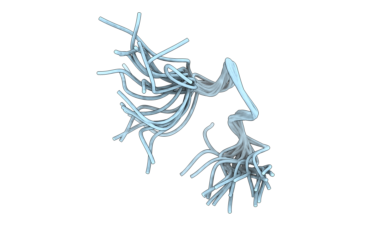
Title:
The third dimensional structure of mab198-bound pep.1 for autoimmune myasthenia gravis
Method Details:
Experimental Method:
Conformers Calculated:
20
Conformers Submitted:
20
Selection Criteria:
structures with the lowest energy