Deposition Date
2006-12-07
Release Date
2007-06-12
Last Version Date
2024-11-20
Entry Detail
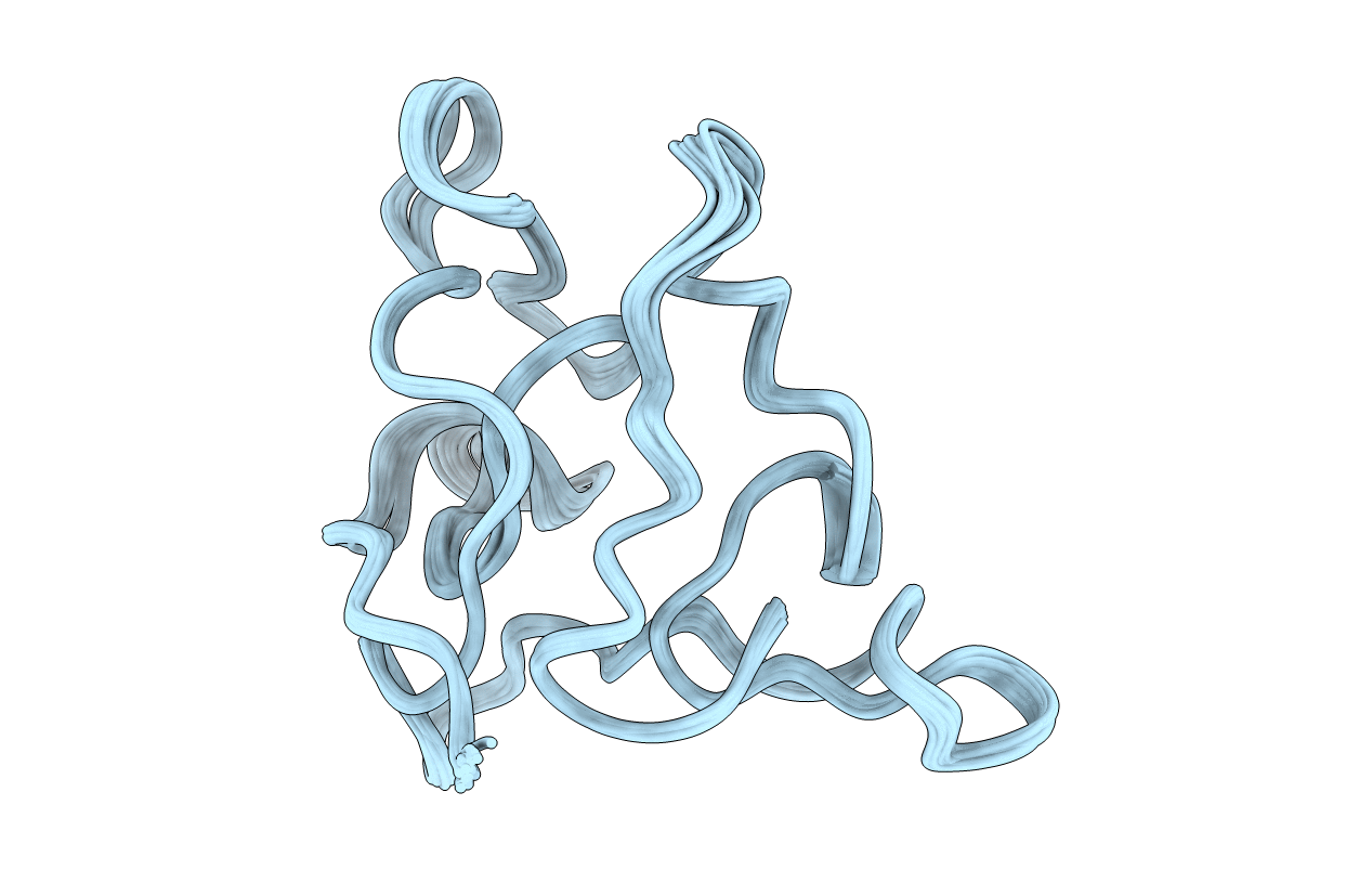
PDB ID:
2JMV
Keywords:
Title:
Solution structure of scytovirin refined against residual dipolar couplings
Biological Source:
Source Organism(s):
Scytonema varium (Taxon ID: 423208)
Expression System(s):
Method Details:
Experimental Method:
Conformers Calculated:
200
Conformers Submitted:
20
Selection Criteria:
structures with the lowest energy