2JJX image
Deposition Date 2008-04-23
Release Date 2008-07-29
Last Version Date 2023-12-13
Entry Detail
PDB ID:
2JJX
Keywords:
Title:
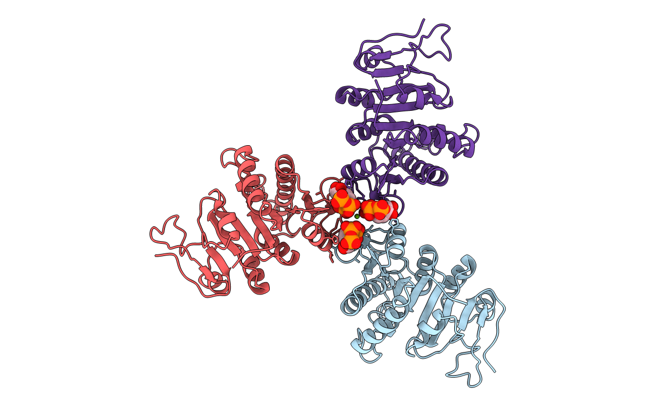
THE CRYSTAL STRUCTURE OF UMP KINASE FROM BACILLUS ANTHRACIS (BA1797)
Biological Source:
Source Organism:
Host Organism:
Method Details:
Experimental Method:
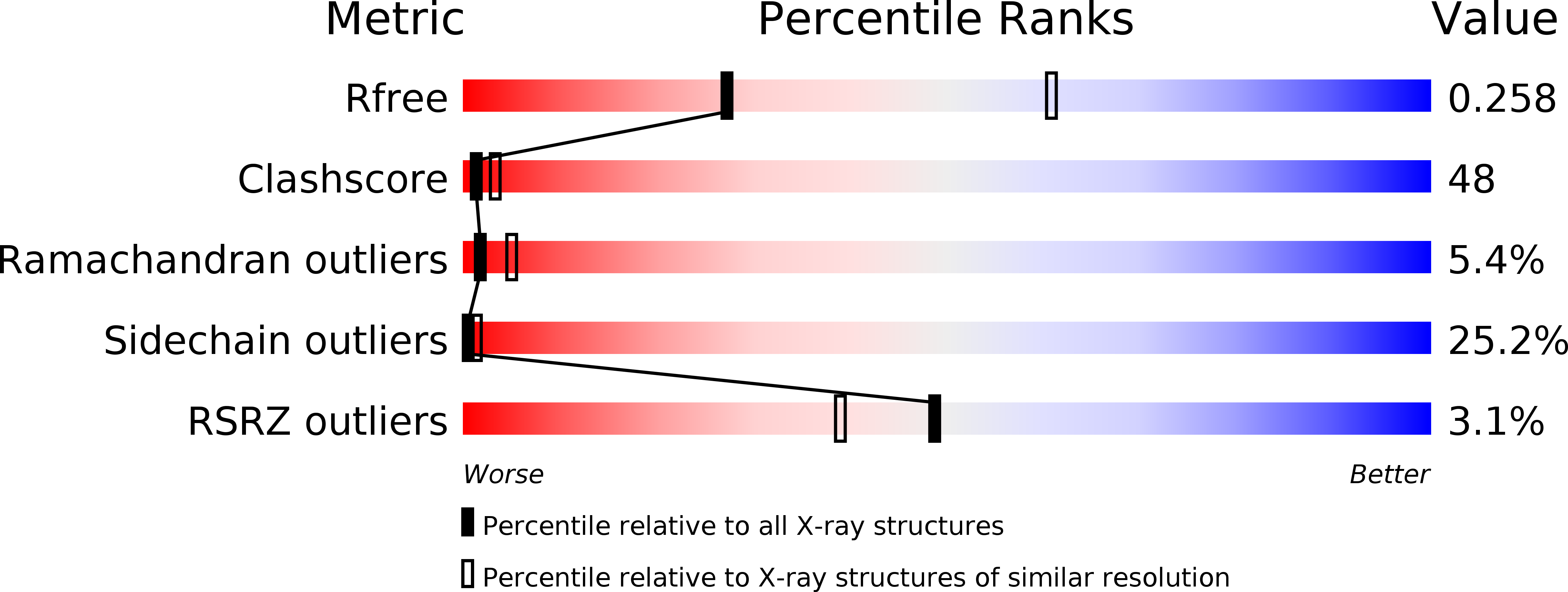
Resolution:
2.82 Å
R-Value Free:
0.25
R-Value Work:
0.20
Space Group:
P 61 2 2
Macromolecular Entities
Structures with similar UniProt ID
Protein Blast
Polymer Type:polypeptide(L)
Molecule:URIDYLATE KINASE
Chain IDs:A, B, C
Chain Length:255
Number of Molecules:3
Biological Source:BACILLUS ANTHRACIS
Primary Citation
The Crystal Structure of Ump Kinase from Bacillus Anthracis (Ba1797) Reveals an Allosteric Nucleotide-Binding Site.
J.Mol.Biol. 381 1098 ? (2008)
PMID: 18625239 DOI: 10.1016/J.JMB.2008.06.078

Abstact

Uridine monophosphate (UMP) kinase is a conserved enzyme that catalyzes the ATP-driven conversion of uridylate monophosphate into uridylate diphosphate, an essential metabolic step. In prokaryotes, the enzyme exists as a homohexamer that is regulated by various metabolites. Whereas the enzymatic mechanism of UMP kinase (UK) is well-characterized, the molecular basis of its regulation remains poorly understood. Here we report the crystal structure of UK from Bacillus anthracis (BA1797) in complex with ATP at 2.82 A resolution. It reveals that the cofactor, in addition to binding in the active sites, also interacts with separate binding pockets located near the center of the hexameric structure. The existence of such an allosteric binding site had been predicted by biochemical studies, but it was not identified in previous crystal structures of prokaryotic UKs. We show that this putative allosteric pocket is conserved across different bacterial species, suggesting that it is a feature common to bacterial UKs, and we present a structural model for the allosteric regulation of this enzyme.

Legend

Protein

Chemical

Disease

Primary Citation of related structures