2JGV image
Deposition Date 2007-02-16
Release Date 2007-04-24
Last Version Date 2024-10-16
Entry Detail
PDB ID:
2JGV
Keywords:
Title:
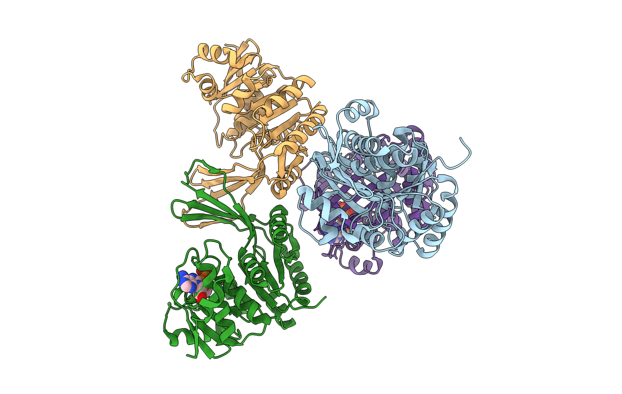
STRUCTURE OF Staphylococcus aureus D-TAGATOSE-6-PHOSPHATE KINASE in complex with ADP
Biological Source:
Source Organism:
Host Organism:
Method Details:
Experimental Method:
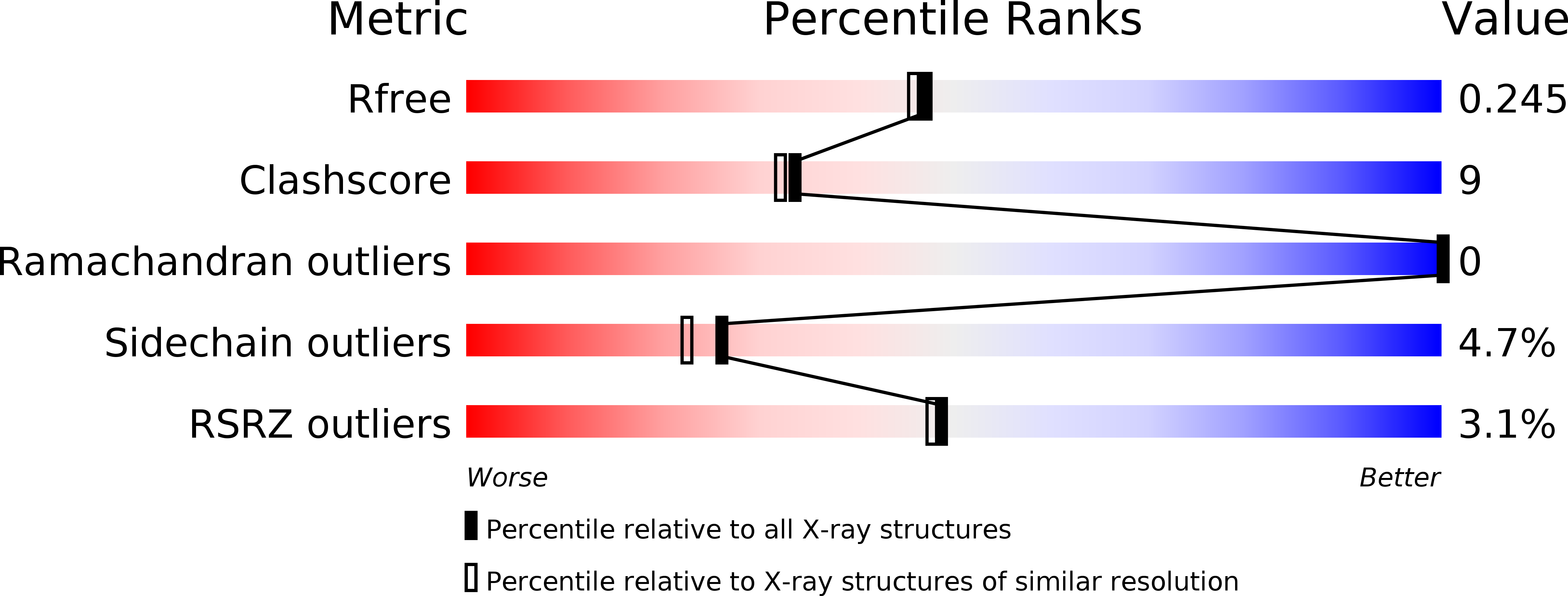
Resolution:
2.00 Å
R-Value Free:
0.24
R-Value Work:
0.19
R-Value Observed:
0.20
Space Group:
P 1 21 1
Macromolecular Entities
Structures with similar UniProt ID
Protein Blast
Polymer Type:polypeptide(L)
Molecule:TAGATOSE-6-PHOSPHATE KINASE
Gene (Uniprot):lacC
Chain IDs:A, B, C, D
Chain Length:330
Number of Molecules:4
Biological Source:STAPHYLOCOCCUS AUREUS
Modified Residue
Compound ID Chain ID Parent Comp ID Details 2D Image
MSE A MET SELENOMETHIONINE
Ligand Molecules
Primary Citation
Structures of Staphylococcus Aureus D-Tagatose-6-Phosphate Kinase Implicate Domain Motions in Specificity and Mechanism.
J.Biol.Chem. 282 19948 ? (2007)
PMID: 17459874 DOI: 10.1074/JBC.M701480200

Abstact

High resolution structures of Staphylococcus aureus d-tagatose-6-phosphate kinase (LacC) in two crystal forms are herein reported. The structures define LacC in apoform, in binary complexes with ADP or the co-factor analogue AMP-PNP, and in a ternary complex with AMP-PNP and D-tagatose-6-phosphate. The tertiary structure of the LacC monomer, which is closely related to other members of the pfkB subfamily of carbohydrate kinases, is composed of a large alpha/beta core domain and a smaller, largely beta "lid." Four extended polypeptide segments connect these two domains. Dimerization of LacC occurs via interactions between lid domains, which come together to form a beta-clasp structure. Residues from both subunits contribute to substrate binding. LacC adopts a closed structure required for phosphoryl transfer only when both substrate and co-factor are bound. A reaction mechanism similar to that used by other phosphoryl transferases is proposed, although unusually, when both substrate and co-factor are bound to the enzyme two Mg(2+) ions are observed in the active site. A new motif of amino acid sequence conservation common to the pfkB subfamily of carbohydrate kinases is identified.

Legend

Protein

Chemical

Disease

Primary Citation of related structures
Feedback Form
Name
Email
Institute
Feedback