Deposition Date
2007-02-14
Release Date
2007-05-01
Last Version Date
2023-12-13
Entry Detail
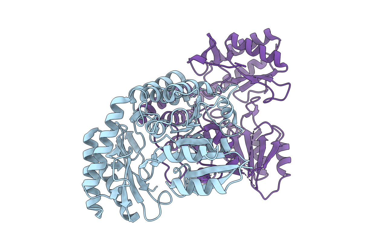
Biological Source:
Source Organism(s):
PSEUDOMONAS PAUCIMOBILIS (Taxon ID: 13689)
Expression System(s):
Method Details:
Experimental Method:
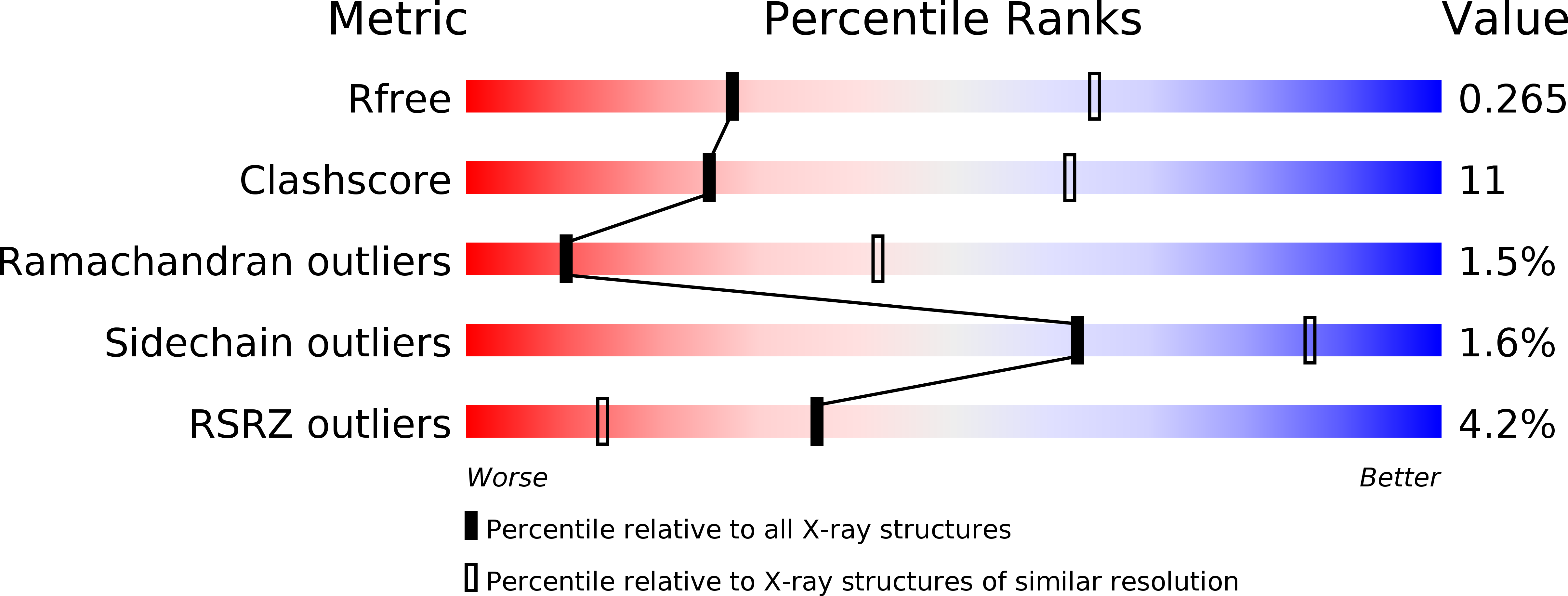
Resolution:
3.00 Å
R-Value Free:
0.27
R-Value Work:
0.25
R-Value Observed:
0.25
Space Group:
C 2 2 21