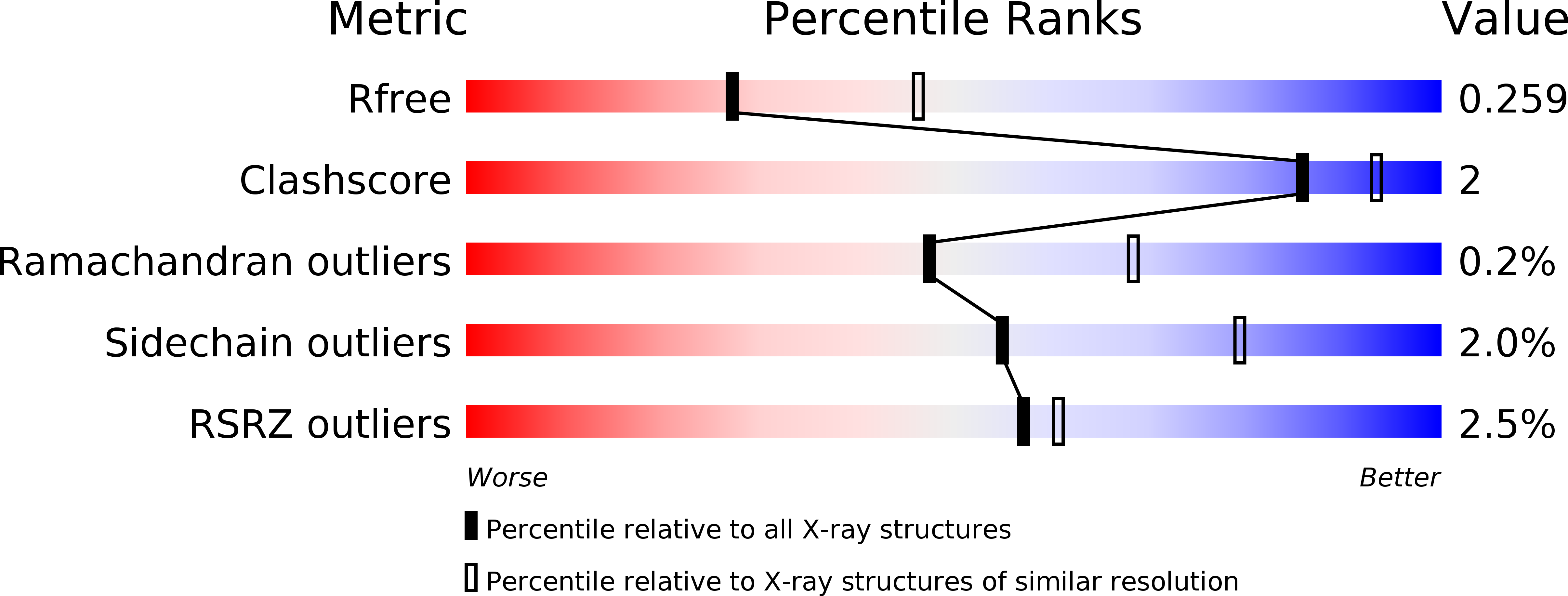
2JFB image
Deposition Date 2007-01-30
Release Date 2007-05-01
Last Version Date 2023-12-13
Entry Detail
PDB ID:
2JFB
Keywords:
Title:
3D Structure of Lumazine Synthase from Candida albicans
Biological Source:
Source Organism:
Host Organism:
Method Details:
Experimental Method:
Resolution:
2.50 Å
R-Value Free:
0.26
R-Value Work:
0.21
R-Value Observed:
0.21
Space Group:
P 31 2 1
Macromolecular Entities
Polymer Type:polypeptide(L)
Molecule:6,7-DIMETHYL-8-RIBITYLLUMAZINE SYNTHASE
Chain IDs:A, B, C, D, E, F, G, H, I, J, K, L, M, N, O
Chain Length:164
Number of Molecules:15
Biological Source:CANDIDA ALBICANS
Primary Citation
Lumazine Synthase from Candida Albicans as an Anti- Fungal Target Enzyme: Structural and Biochemical Basis for Drug Design.
J.Biol.Chem. 282 17231 ? (2007)
PMID: 17446177 DOI: 10.1074/JBC.M701724200

Abstact

Lumazine synthase is an enzyme involved in riboflavin biosynthesis in many plants and microorganisms, including numerous human pathogens. The fact that the enzymes of the riboflavin biosynthesis pathway are not present in the human or animal host makes them potential targets for anti-infective agents. The crystal structure of lumazine synthase from Candida albicans was solved by molecular replacement and refined at 2.5-Angstrom resolution. The results of crystallographic investigations and sedimentation equilibrium experiments clearly indicated the presence of pentameric assemblies of the enzyme either in crystals or in solution. Isothermal titration calorimetry measurements of the binding reactions of four different inhibitors revealed high affinity for all four compounds with binding constants in the micromolar range. Structural comparison with previously determined structures of the enzyme.ligand complexes of other orthologue allowed modeling of the binding of four different inhibitors into the active site of lumazine synthase from Candida albicans.

Legend

Protein

Chemical

Disease

Primary Citation of related structures