2JET image
Deposition Date 2007-01-22
Release Date 2007-09-18
Last Version Date 2024-11-06
Entry Detail
PDB ID:
2JET
Keywords:
Title:
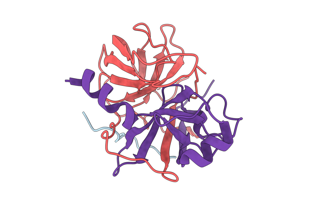
Crystal structure of a trypsin-like mutant (S189D , A226G) chymotrypsin.
Biological Source:
Source Organism:
Host Organism:
Method Details:
Experimental Method:
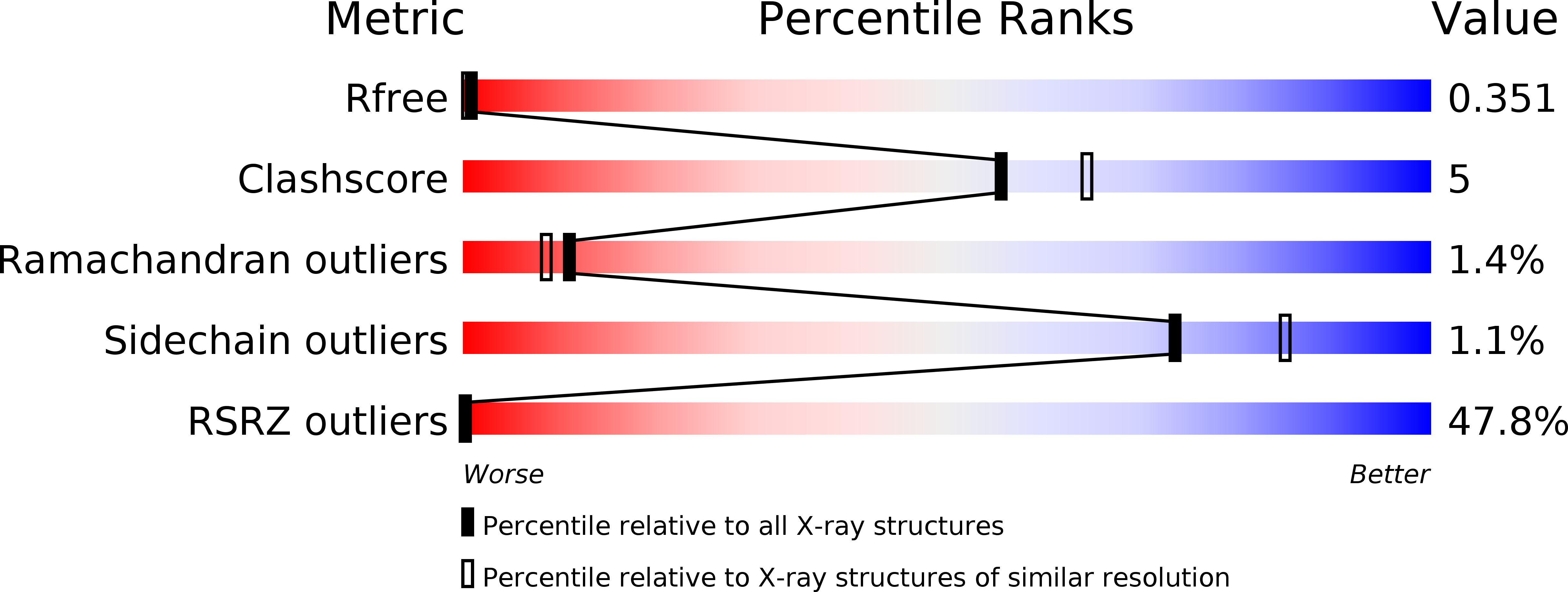
Resolution:
2.20 Å
R-Value Free:
0.33
R-Value Work:
0.27
R-Value Observed:
0.27
Space Group:
P 1 21 1
Macromolecular Entities
Polymer Type:polypeptide(L)
Molecule:CHYMOTRYPSINOGEN B CHAIN A
Gene (Uniprot):Ctrb1
Chain IDs:A
Chain Length:15
Number of Molecules:1
Biological Source:RATTUS NORVEGICUS
Polymer Type:polypeptide(L)
Molecule:CHYMOTRYPSINOGEN B CHAIN B
Gene (Uniprot):Ctrb1
Chain IDs:B
Chain Length:128
Number of Molecules:1
Biological Source:RATTUS NORVEGICUS
Polymer Type:polypeptide(L)
Molecule:CHYMOTRYPSINOGEN B CHAIN C
Gene (Uniprot):Ctrb1
Mutations:YES
Chain IDs:C
Chain Length:99
Number of Molecules:1
Biological Source:RATTUS NORVEGICUS
Primary Citation
The Crystal Structure of a Trypsin-Like Mutant Chymotrypsin: The Role of Position 226 in the Activity and Specificity of S189D Chymotrypsin.
Protein J. 27 79 ? (2008)
PMID: 17805946 DOI: 10.1007/S10930-007-9110-3

Abstact

The crystal structure of the S189D+A226G rat chymotrypsin-B mutant has been determined at 2.2 angstroms resolution. This mutant is the most trypsin-like mutant so far in the line of chymotrypsin-to-trypsin conversions, aiming for a more complete understanding of the structural basis of substrate specificity in pancreatic serine proteases. A226G caused significant rearrangements relative to S189D chymotrypsin, allowing an internal conformation of Asp189 which is close to that in trypsin. Serious distortions remain, however, in the activation domain, including zymogen-like features. The pH-profile of activity suggests that the conformation of the S1-site of the mutant is influenced also by the P1 residue of the substrate.

Legend

Protein

Chemical

Disease

Primary Citation of related structures