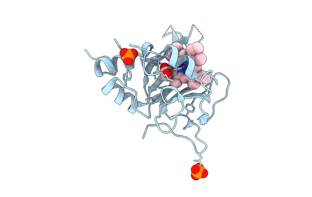
2JE3 image
Entry Detail
PDB ID:
2JE3
Title:
Cytochrome P460 from Nitrosomonas europaea - probable physiological form
Biological Source:
Source Organism:
Host Organism:
PDB Version:
Deposition Date:
2007-01-13
Release Date:
2007-07-03
Method Details:
Experimental Method:
Resolution:
1.80 Å
R-Value Free:
0.23
R-Value Work:
0.19
R-Value Observed:
0.19
Space Group:
P 31 2 1
Macromolecular Entities
Polymer Type:polypeptide(L)
Description:CYTOCHROME P460
Chain IDs:A
Chain Length:186
Number of Molecules:1
Biological Source:NITROSOMONAS EUROPAEA
Primary Citation
The Crystal Structure of Cytochrome P460 of Nitrosomonas Europaea Reveals a Novel Cytochrome Fold and Heme-Protein Cross-Link.
Biochemistry 46 8340 ? (2007)
PMID: 17583915 DOI: 10.1021/BI700086R

Abstact

We have determined the 1.8 A X-ray crystal structure of a monoheme c-type cytochrome, cytochrome P460, from Nitrosomonas europea. The chromophore possesses unusual spectral properties analogous to those of the catalytic heme P460 of hydroxylamine oxidoreductase (HAO), the only known heme in biology to withdraw electrons from an iron-coordinated substrate. The analysis reveals a homodimeric structure and elucidates a new c-type cytochrome fold that is predominantly beta-sheet. In addition to the two cysteine thioether links to the porphyrin typical of c-type hemes, there is a third proteinaceous link involving a conserved lysine. The covalent bond is between the lysine side-chain nitrogen and the 13'-meso carbon of the heme, which, following cross-link formation, is sp3-hybridized, demonstrating the loss of conjugation at this position within the porphyrin. The structure has implications for the analogous tyrosine-heme meso carbon cross-link observed in HAO.

Legend

Protein

Chemical

Disease

Primary Citation of related structures