2JDI image
Deposition Date 2007-01-09
Release Date 2007-03-13
Last Version Date 2023-12-13
Entry Detail
PDB ID:
2JDI
Keywords:
Title:
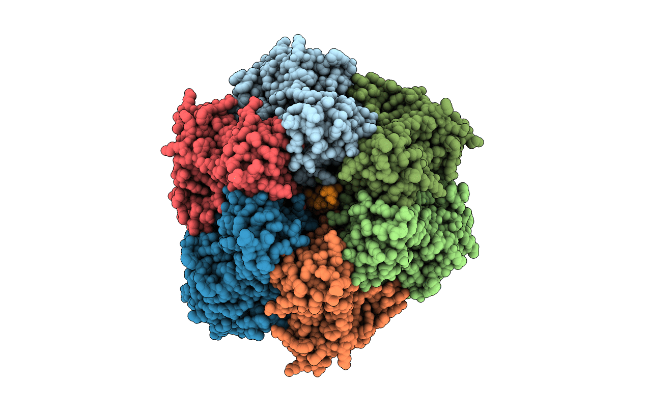
Ground state structure of F1-ATPase from bovine heart mitochondria (Bovine F1-ATPase crystallised in the absence of azide)
Biological Source:
Source Organism:
BOS TAURUS (Taxon ID: 9913)
Method Details:
Experimental Method:
Resolution:
1.90 Å
R-Value Free:
0.22
R-Value Work:
0.17
R-Value Observed:
0.17
Space Group:
P 21 21 21
Macromolecular Entities
Polymer Type:polypeptide(L)
Molecule:ATP SYNTHASE SUBUNIT ALPHA HEART ISOFORM
Gene (Uniprot):ATP5F1A
Chain IDs:A, B, C
Chain Length:510
Number of Molecules:3
Biological Source:BOS TAURUS
Polymer Type:polypeptide(L)
Molecule:ATP SYNTHASE SUBUNIT BETA
Gene (Uniprot):ATP5F1B
Chain IDs:D, E, F
Chain Length:482
Number of Molecules:3
Biological Source:BOS TAURUS
Polymer Type:polypeptide(L)
Molecule:ATP SYNTHASE GAMMA CHAIN
Gene (Uniprot):ATP5F1C
Chain IDs:G
Chain Length:273
Number of Molecules:1
Biological Source:BOS TAURUS
Polymer Type:polypeptide(L)
Molecule:ATP SYNTHASE DELTA CHAIN
Gene (Uniprot):ATP5F1D
Chain IDs:H
Chain Length:146
Number of Molecules:1
Biological Source:BOS TAURUS
Polymer Type:polypeptide(L)
Molecule:ATP SYNTHASE EPSILON CHAIN
Gene (Uniprot):ATP5F1E
Chain IDs:I
Chain Length:50
Number of Molecules:1
Biological Source:BOS TAURUS
Primary Citation
Ground State Structure of F1-ATPase from Bovine Heart Mitochondria at 1.9 A Resolution
J.Biol.Chem. 282 14238 ? (2007)
PMID: 17350959 DOI: 10.1074/JBC.M700203200

Abstact

The structure of bovine F(1)-ATPase, crystallized in the presence of AMP-PNP and ADP, but in the absence of azide, has been determined at 1.9A resolution. This structure has been compared with the previously described structure of bovine F(1)-ATPase determined at 1.95A resolution with crystals grown under the same conditions but in the presence of azide. The two structures are extremely similar, but they differ in the nucleotides that are bound to the catalytic site in the beta(DP)-subunit. In the present structure, the nucleotide binding sites in the beta(DP)- and beta(TP)-subunits are both occupied by AMP-PNP, whereas in the earlier structure, the beta(TP) site was occupied by AMP-PNP and the beta(DP) site by ADP, where its binding is enhanced by a bound azide ion. Also, the conformation of the side chain of the catalytically important residue, alphaArg-373 differs in the beta(DP)- and beta(TP)-subunits. Thus, the structure with bound azide represents the ADP inhibited state of the enzyme, and the new structure represents a ground state intermediate in the active catalytic cycle of ATP hydrolysis.

Legend

Protein

Chemical

Disease

Primary Citation of related structures