2JD6 image
Entry Detail
PDB ID:
2JD6
Keywords:
Title:
Crystal Structure of the as isolated Ferritin from the Hyperthermophilic Archaeal Anaerobe Pyrococcus furiosus
Biological Source:
Source Organism:
Host Organism:
PDB Version:
Deposition Date:
2007-01-05
Release Date:
2007-02-27
Method Details:
Experimental Method:
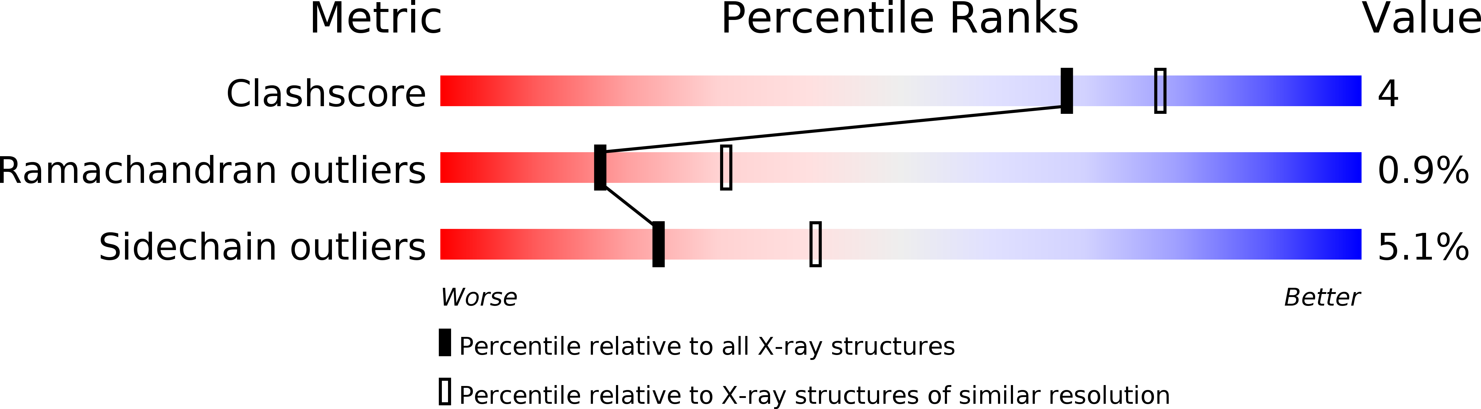
Resolution:
2.75 Å
R-Value Free:
0.24
R-Value Work:
0.19
R-Value Observed:
0.19
Space Group:
C 2 2 21
Macromolecular Entities
Polymer Type:polypeptide(L)
Description:FERRITIN HOMOLOG
Chain IDs:A (auth: 0), B (auth: 1), C (auth: 2), D (auth: 3), E (auth: 4), F (auth: 5), G (auth: 6), H (auth: 7), I (auth: 8), J (auth: 9), K (auth: A), L (auth: B), M (auth: C), N (auth: D), O (auth: E), P (auth: F), Q (auth: G), R (auth: H), S (auth: I), T (auth: J), U (auth: K), V (auth: L), W (auth: M), X (auth: N), Y (auth: O), Z (auth: P), AA (auth: Q), BA (auth: R), CA (auth: S), DA (auth: T), EA (auth: U), FA (auth: V), GA (auth: W), HA (auth: X), IA (auth: Y), JA (auth: Z)
Chain Length:174
Number of Molecules:36
Biological Source:PYROCOCCUS FURIOSUS
Primary Citation
Crystal Structure of the Ferritin from the Hyperthermophilic Archaeal Anaerobe Pyrococcus Furiosus
J.Biol.Inorg.Chem. 12 615 ? (2007)
PMID: 17541801 DOI: 10.1007/S00775-007-0212-3

Abstact

The crystal structure of the ferritin from the archaeon, hyperthermophile and anaerobe Pyrococcus furiosus (PfFtn) is presented. While many ferritin structures from bacteria to mammals have been reported, until now only one was available from archaea, the ferritin from Archaeoglobus fulgidus (AfFtn). The PfFtn 24-mer exhibits the 432 point-group symmetry that is characteristic of most ferritins, which suggests that the 23 symmetry found in the previously reported AfFtn is not a common feature of archaeal ferritins. Consequently, the four large pores that were found in AfFtn are not present in PfFtn. The structure has been solved by molecular replacement and refined at 2.75-Angstrom resolution to R = 0.195 and R(free) = 0.247. The ferroxidase center of the aerobically crystallized ferritin contains one iron at site A and shows sites B and C only upon iron or zinc soaking. Electron paramagnetic resonance studies suggest this iron depletion of the native ferroxidase center to be a result of a complexation of iron by the crystallization salt. The extreme thermostability of PfFtn is compared with that of eight structurally similar ferritins and is proposed to originate mostly from the observed high number of intrasubunit hydrogen bonds. A preservation of the monomer fold, rather than the 24-mer assembly, appears to be the most important factor that protects the ferritin from inactivation by heat.

Legend

Protein

Chemical

Disease

Primary Citation of related structures