2JCM image
Deposition Date 2006-12-27
Release Date 2007-01-30
Last Version Date 2024-11-13
Entry Detail
PDB ID:
2JCM
Keywords:
Title:
Crystal structure of Human Cytosolic 5'-Nucleotidase II in complex with beryllium trifluoride
Biological Source:
Source Organism:
HOMO SAPIENS (Taxon ID: 9606)
Host Organism:
Method Details:
Experimental Method:
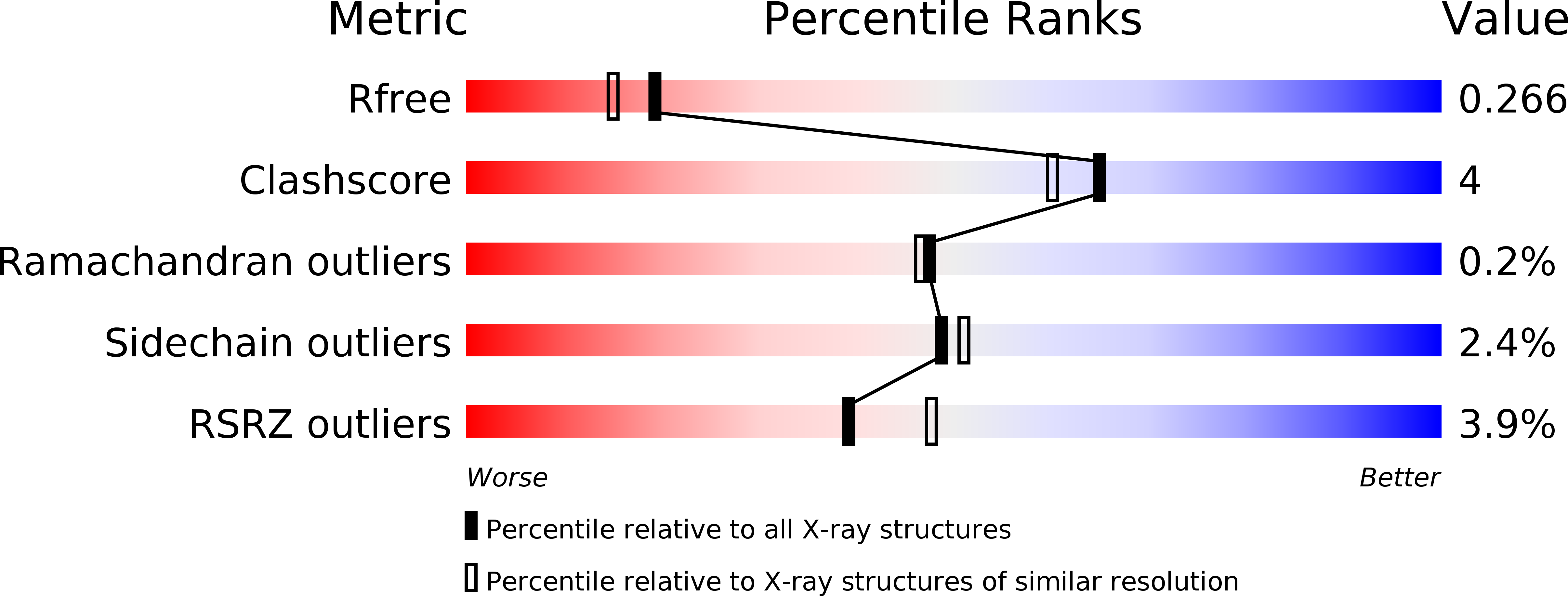
Resolution:
2.15 Å
R-Value Free:
0.26
R-Value Work:
0.21
R-Value Observed:
0.21
Space Group:
I 2 2 2
Macromolecular Entities
Polymer Type:polypeptide(L)
Molecule:CYTOSOLIC PURINE 5'-NUCLEOTIDASE
Gene (Uniprot):NT5C2
Chain IDs:A
Chain Length:555
Number of Molecules:1
Biological Source:HOMO SAPIENS
Modified Residue
Compound ID Chain ID Parent Comp ID Details 2D Image
BFD A ASP ASPARTATE BERYLLIUM TRIFLUORIDE
Primary Citation
Crystal Structure of Human Cytosolic 5'- Nucleotidase II: Insights Into Allosteric Regulation and Substrate Recognition
J.Biol.Chem. 282 17828 ? (2007)
PMID: 17405878 DOI: 10.1074/JBC.M700917200

Abstact

Cytosolic 5'-nucleotidase II catalyzes the dephosphorylation of 6-hydroxypurine nucleoside 5'-monophosphates and regulates the IMP and GMP pools within the cell. It possesses phosphotransferase activity and thereby also catalyzes the reverse reaction. Both reactions are allosterically activated by adenine-based nucleotides and 2,3-bisphosphoglycerate. We have solved structures of cytosolic 5'-nucleotidase II as native protein (2.2 Angstrom) and in complex with adenosine (1.5 Angstrom) and beryllium trifluoride (2.15 Angstrom) The tetrameric enzyme is structurally similar to enzymes of the haloacid dehalogenase (HAD) superfamily, including mitochondrial 5'(3')-deoxyribonucleotidase and cytosolic 5'-nucleotidase III but possesses additional regulatory regions that contain two allosteric effector sites. At effector site 1 located near a subunit interface we modeled diadenosine tetraphosphate with one adenosine moiety in each subunit. This efficiently glues the tetramer subunits together in pairs. The model shows why diadenosine tetraphosphate but not diadenosine triphosphate activates the enzyme and supports a role for cN-II during apoptosis when the level of diadenosine tetraphosphate increases. We have also modeled 2,3-bisphosphoglycerate in effector site 1 using one phosphate site from each subunit. By comparing the structure of cytosolic 5'-nucleotidase II with that of mitochondrial 5'(3')-deoxyribonucleotidase in complex with dGMP, we identified residues involved in substrate recognition.

Legend

Protein

Chemical

Disease

Primary Citation of related structures