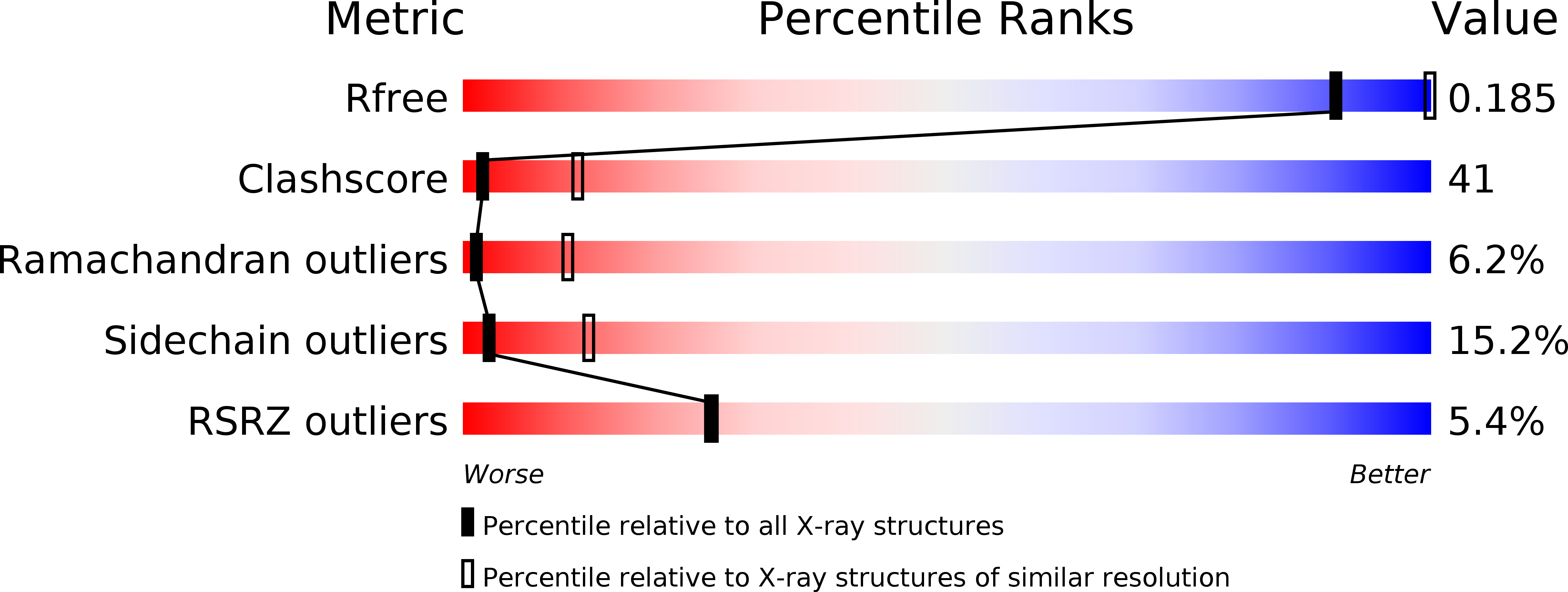
2JBP image
Deposition Date 2006-12-09
Release Date 2007-03-20
Last Version Date 2023-12-13
Entry Detail
PDB ID:
2JBP
Keywords:
Title:
Protein kinase MK2 in complex with an inhibitor (crystal form-2, co- crystallization)
Biological Source:
Source Organism:
HOMO SAPIENS (Taxon ID: 9606)
Host Organism:
Method Details:
Experimental Method:
Resolution:
3.31 Å
R-Value Free:
0.27
R-Value Work:
0.21
Space Group:
P 21 21 21
Macromolecular Entities
Structures with similar UniProt ID
Protein Blast
Polymer Type:polypeptide(L)
Molecule:MAP KINASE-ACTIVATED PROTEIN KINASE 2
Gene (Uniprot):MAPKAPK2
Chain IDs:A, B, C, D, E, F, G, H, I, J, K, L
Chain Length:326
Number of Molecules:12
Biological Source:HOMO SAPIENS
Ligand Molecules
Primary Citation
Structural basis for a high affinity inhibitor bound to protein kinase MK2.
J. Mol. Biol. 369 735 745 (2007)
PMID: 17449059 DOI: 10.1016/j.jmb.2007.03.004

Abstact

The Ser/Thr protein kinase MAPKAP kinase 2 (MK2) plays a crucial role in inflammation. We determined the structure of the kinase domain of MK2 in complex with a low molecular mass inhibitor in two different crystal forms, obtained from soaking and co-crystallization. To our knowledge, these are the first structures of MK2 showing the binding mode of an inhibitor with high binding affinity (IC50 8.5 nM). The two crystal forms revealed conformational flexibility in the binding site and extend the experimental basis for rational drug design. Crystal form-1 contained one MK2 molecule per asymmetric unit. Form-2 contained 12 molecules, which arrange into two different types of MK2 trimers. One of them may serve as a model for an intermediate state during substrate phosphorylation, as each MK2 monomer places its activation segment into the substrate peptide binding groove of the trimer neighbor.

Legend

Protein

Chemical

Disease

Primary Citation of related structures