2JB9 image
Deposition Date 2006-12-05
Release Date 2007-01-04
Last Version Date 2023-12-13
Entry Detail
PDB ID:
2JB9
Keywords:
Title:
PhoB response regulator receiver domain constitutively-active double mutant D10A and D53E.
Biological Source:
Source Organism:
ESCHERICHIA COLI (Taxon ID: 562)
Host Organism:
Method Details:
Experimental Method:
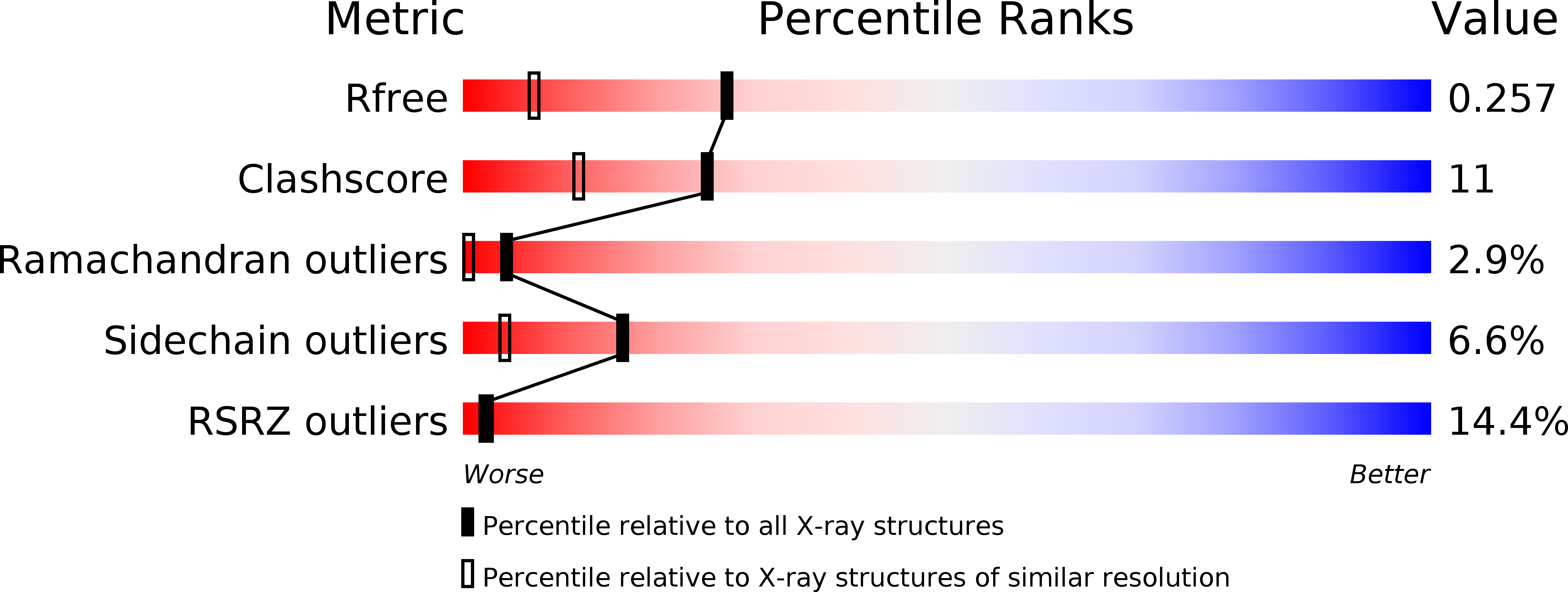
Resolution:
1.70 Å
R-Value Free:
0.25
R-Value Work:
0.22
R-Value Observed:
0.22
Space Group:
P 21 21 21
Macromolecular Entities
Polymer Type:polypeptide(L)
Molecule:PHOSPHATE REGULON TRANSCRIPTIONAL REGULATORY PROTEIN PHOB
Gene (Uniprot):phoB
Mutations:YES
Chain IDs:A, B
Chain Length:127
Number of Molecules:2
Biological Source:ESCHERICHIA COLI
Primary Citation
The X-Ray Crystal Structures of Two Constitutively Active Mutants of the E. Coli Phob Receiver Domain Give Insights Into Activation
J.Mol.Biol. 366 626 ? (2007)
PMID: 17182055 DOI: 10.1016/J.JMB.2006.11.038

Abstact

The PhoR/PhoB two-component system is a key regulatory protein network enabling Escherichia coli to respond to inorganic phosphate (Pi) starvation conditions by turning on Pho regulon genes for more efficient Pi uptake and use of alternative phosphorus sources. Under environmental Pi depletion, the response regulator (RR) component, PhoB, is phosphorylated at the receiver domain (RD), a process that requires Mg(2+) bound at the active site. Phosphorylation of the RD relieves the inhibition of the PhoB effector domain (ED), a DNA-binding region that binds to Pho regulon promoters to activate transcription. The molecular details of the activation are proposed to involve dimerization of the RD and a conformational change in the RD detected by the ED. The structure of the PhoB RD shows a symmetrical interaction involving alpha1, loop beta5alpha5 and N terminus of alpha5 elements, also seen in the complex of PhoB RD with Mg(2+), in which helix alpha4 highly increases its flexibility. PhoB RD in complex with Mg(2+) and BeF(3) (an emulator of the phosphate moiety) undergoes a dramatic conformational change on helix alpha4 and shows another interaction involving alpha4, beta5 and alpha5 segments. We have selected a series of constitutively active PhoB mutants (PhoB(CA)) that are able to turn on the Pho regulon promoters in the absence phosphorylation and, as they cannot be inactivated, should therefore mimic the active RD state of PhoB and its functional oligomerisation. We have analysed the PhoB(CA) RD crystal structures of two such mutants, Asp53Ala/Tyr102Cys and Asp10Ala/Asp53Glu. Interestingly, both mutants reproduce the homodimeric arrangement through the symmetric interface encountered in the unbound and magnesium-bound wild-type PhoB RD structures. Besides, the mutant RD structures show a modified active site organization as well as changes at helix alpha4 that correlate with repositioning of surrounding residues, like the active-site events indicator Trp54, putatively redifining the interaction with the ED in the full-length protein.

Legend

Protein

Chemical

Disease

Primary Citation of related structures