2JB0 image
Deposition Date 2006-12-01
Release Date 2007-04-03
Last Version Date 2023-12-13
Entry Detail
PDB ID:
2JB0
Title:
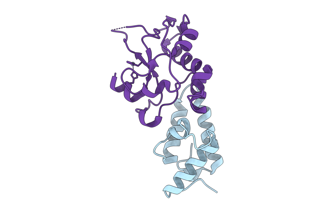
CRYSTAL STRUCTURE OF THE MUTANT H573A OF THE NUCLEASE DOMAIN OF COLE7 IN COMPLEX WITH IM7
Biological Source:
Source Organism:
Host Organism:
Method Details:
Experimental Method:
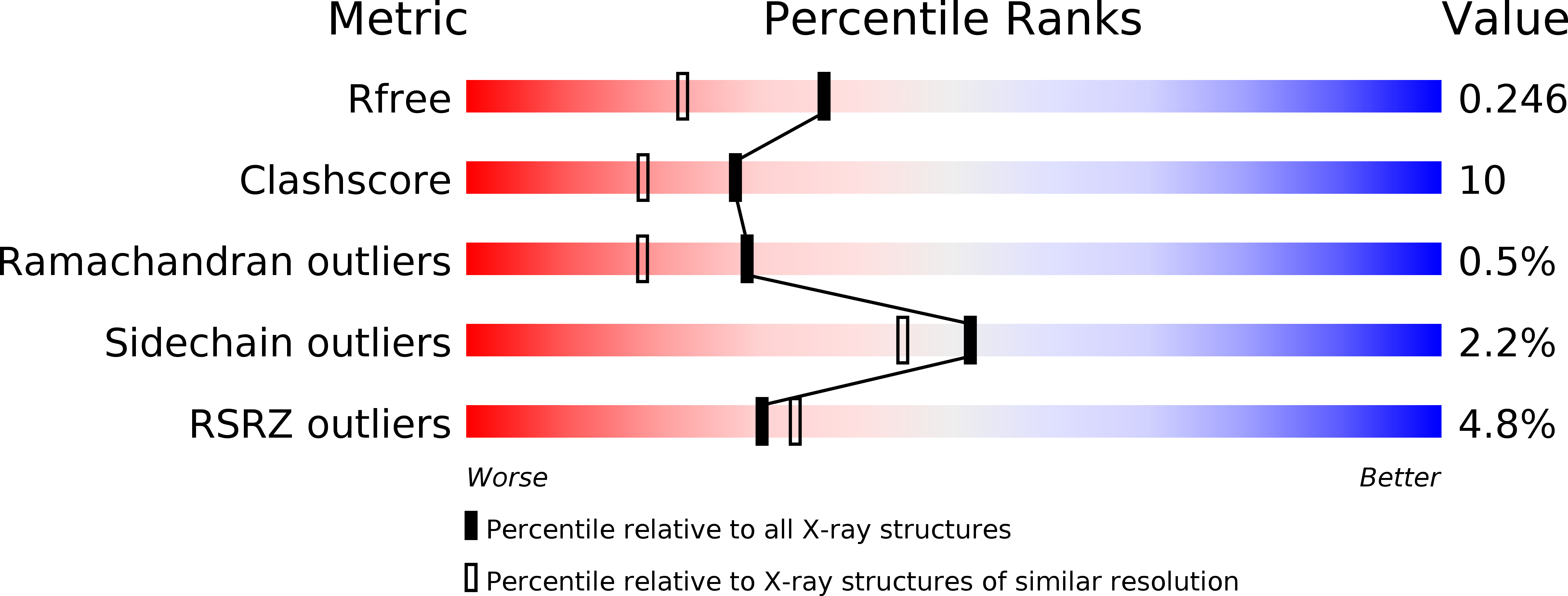
Resolution:
1.91 Å
R-Value Free:
0.25
R-Value Work:
0.21
R-Value Observed:
0.21
Space Group:
I 2 2 2
Macromolecular Entities
Polymer Type:polypeptide(L)
Molecule:COLICIN E7 IMMUNITY PROTEIN
Gene (Uniprot):imm
Chain IDs:A
Chain Length:87
Number of Molecules:1
Biological Source:ESCHERICHIA COLI
Polymer Type:polypeptide(L)
Molecule:COLICIN E7
Gene (Uniprot):colE7
Mutations:YES
Chain IDs:B
Chain Length:131
Number of Molecules:1
Biological Source:ESCHERICHIA COLI
Ligand Molecules
Primary Citation
The Conserved Asparagine in the Hnh Motif Serves an Important Structural Role in Metal Finger Endonucleases.
J.Mol.Biol. 368 812 ? (2007)
PMID: 17368670 DOI: 10.1016/J.JMB.2007.02.044

Abstact

The HNH motif is a small nucleic acid binding and cleavage module, widespread in metal finger endonucleases in all life kingdoms. Here we studied a non-specific endonuclease, the nuclease domain of ColE7 (N-ColE7), to decipher the role of the conserved asparagine and histidine residues in the HNH motif. We found, using fluorescence resonance energy transfer (FRET) assays, that the DNA hydrolysis activity of H545 N-ColE7 mutants was completely abolished while activities of N560 and H573 mutants varied from 6.9% to 83.2% of the wild-type activity. The crystal structures of three N-ColE7 mutants in complex with the inhibitor Im7, N560A-Im7, N560D-Im7 and H573A-Im7, were determined at a resolution of 1.9 A to 2.2 A. H573 is responsible for metal ion binding in the wild-type protein, as the zinc ion is still partially associated in the structure of H573A, suggesting that H573 plays a supportive role in metal binding. Both N560A and N560D contain a disordered loop in the HNH motif due to the disruption of the hydrogen bond network surrounding the side-chain of residue 560, and as a result, the imidazole ring of the general base residue H545 is tilted slightly and the scissile phosphate is shifted, leading to the large reductions in hydrolysis activities. These results suggest that the highly conserved asparagine in the HNH motif, in general, plays a structural role in constraining the loop in the metal finger structure and keeping the general base histidine and scissile phosphate in the correct position for DNA hydrolysis.

Legend

Protein

Chemical

Disease

Primary Citation of related structures