
Deposition Date
2006-10-31
Release Date
2006-11-07
Last Version Date
2024-11-06
Entry Detail
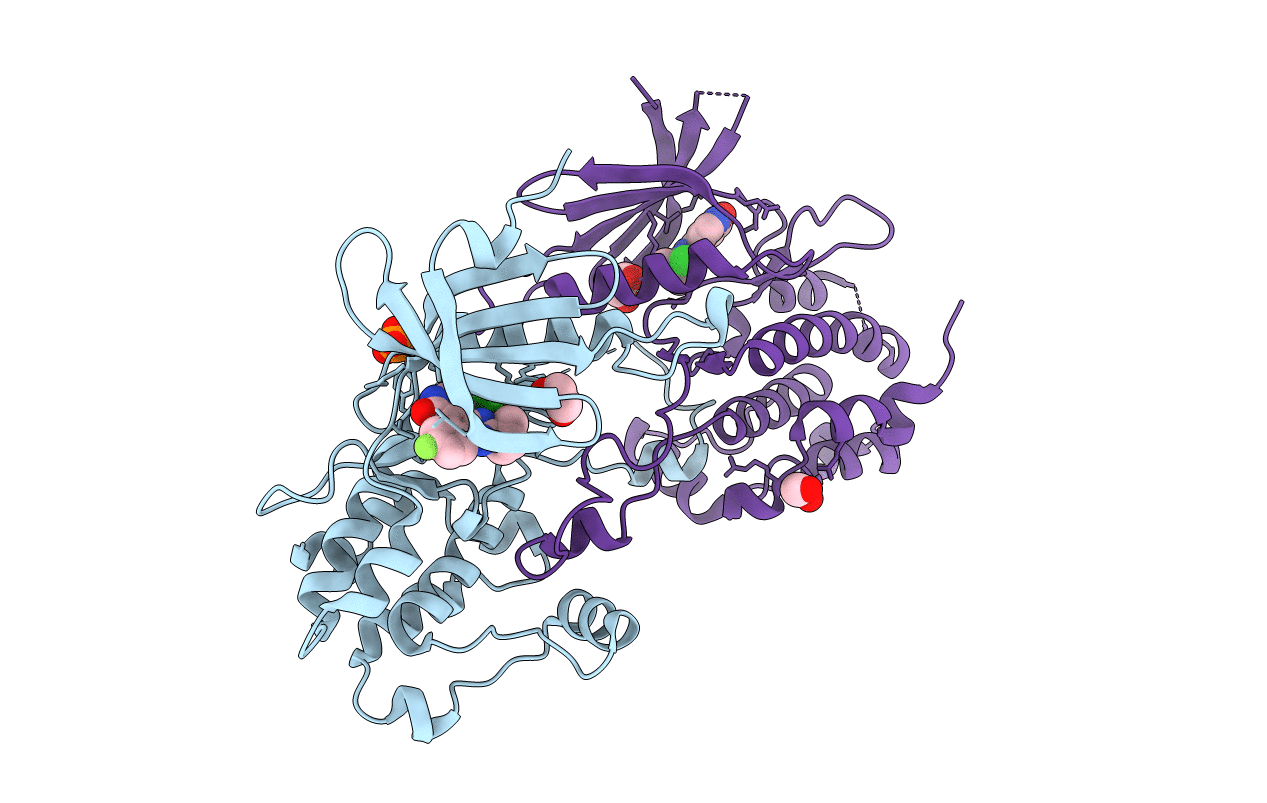
PDB ID:
2J90
Keywords:
Title:
Crystal structure of human ZIP kinase in complex with a tetracyclic pyridone inhibitor (Pyridone 6)
Biological Source:
Source Organism(s):
HOMO SAPIENS (Taxon ID: 9606)
Expression System(s):
Method Details:
Experimental Method:
Resolution:
2.00 Å
R-Value Free:
0.22
R-Value Work:
0.18
R-Value Observed:
0.18
Space Group:
P 43 21 2
