Deposition Date
2006-10-31
Release Date
2006-12-13
Last Version Date
2023-12-13
Entry Detail
PDB ID:
2J8X
Keywords:
Title:
Epstein-Barr virus uracil-DNA glycosylase in complex with Ugi from PBS-2
Biological Source:
Source Organism(s):
EPSTEIN-BARR VIRUS (Taxon ID: 10376)
BACILLUS PHAGE PBS2 (Taxon ID: 10684)
BACILLUS PHAGE PBS2 (Taxon ID: 10684)
Expression System(s):
Method Details:
Experimental Method:
Resolution:
2.30 Å
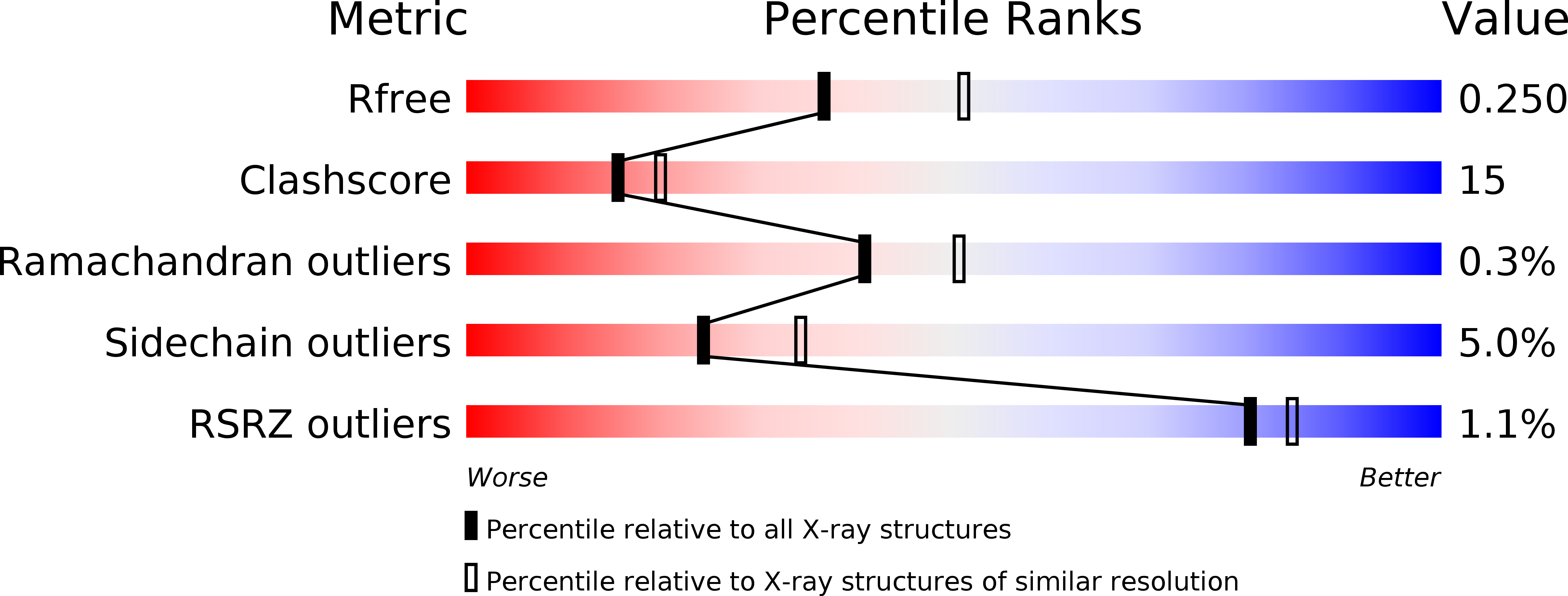
R-Value Free:
0.25
R-Value Work:
0.19
R-Value Observed:
0.19
Space Group:
C 2 2 21