2J6V image
Deposition Date 2006-10-04
Release Date 2007-10-16
Last Version Date 2024-10-23
Entry Detail
PDB ID:
2J6V
Keywords:
Title:
Crystal structure of the DNA repair enzyme UV Damage Endonuclease
Biological Source:
Source Organism:
Host Organism:
Method Details:
Experimental Method:
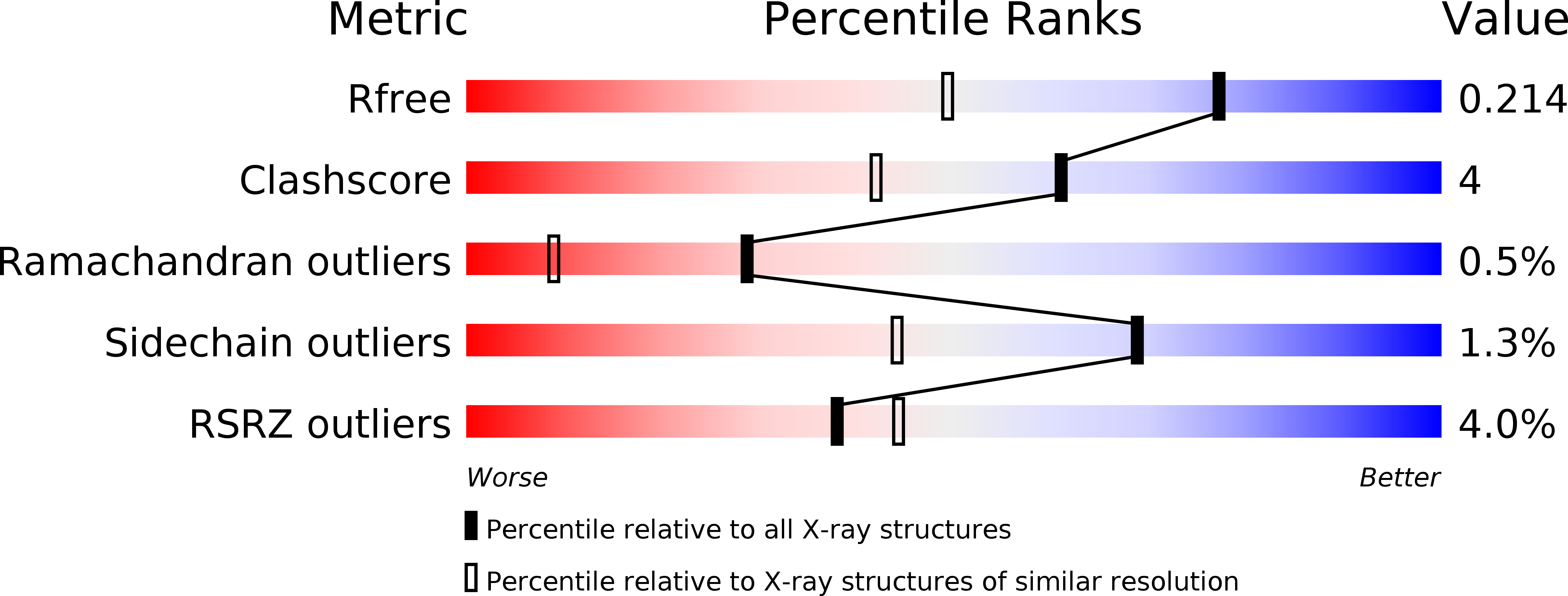
Resolution:
1.55 Å
R-Value Free:
0.21
R-Value Work:
0.18
R-Value Observed:
0.18
Space Group:
P 1
Macromolecular Entities
Structures with similar UniProt ID
Protein Blast
Polymer Type:polypeptide(L)
Molecule:UV ENDONUCLEASE
Gene (Uniprot):TT_P0052
Chain IDs:A, B
Chain Length:301
Number of Molecules:2
Biological Source:THERMUS THERMOPHILUS
Modified Residue
Compound ID Chain ID Parent Comp ID Details 2D Image
ALY A LYS N(6)-ACETYLLYSINE
KCX A LYS LYSINE NZ-CARBOXYLIC ACID
MSE A MET SELENOMETHIONINE
Primary Citation
Crystal Structure of the DNA Repair Enzyme Ultraviolet Damage Endonuclease.
Structure 15 1316 ? (2007)
PMID: 17937920 DOI: 10.1016/J.STR.2007.05.010

Abstact

The ultraviolet damage endonuclease (UVDE) performs the initial step in an alternative excision repair pathway of UV-induced DNA damage, nicking immediately adjacent to the 5' phosphate of the damaged nucleotides. Unique for a single-protein DNA repair endonuclease, it can detect different types of damage. Here we show that Thermus thermophilus UVDE shares some essential structural features with Endo IV, an enzyme from the base excision repair pathway that exclusively nicks at abasic sites. A comparison between the structures indicates how DNA is bound by UVDE, how UVDE may recognize damage, and which of its residues are involved in catalysis. Furthermore, the comparison suggests an elegant explanation of UVDE's potential to recognize different types of damage. Incision assays including point mutants of UVDE confirmed the relevance of these conclusions.

Legend

Protein

Chemical

Disease

Primary Citation of related structures