2J5M image
Deposition Date 2006-09-18
Release Date 2006-12-06
Last Version Date 2024-11-13
Entry Detail
PDB ID:
2J5M
Keywords:
Title:
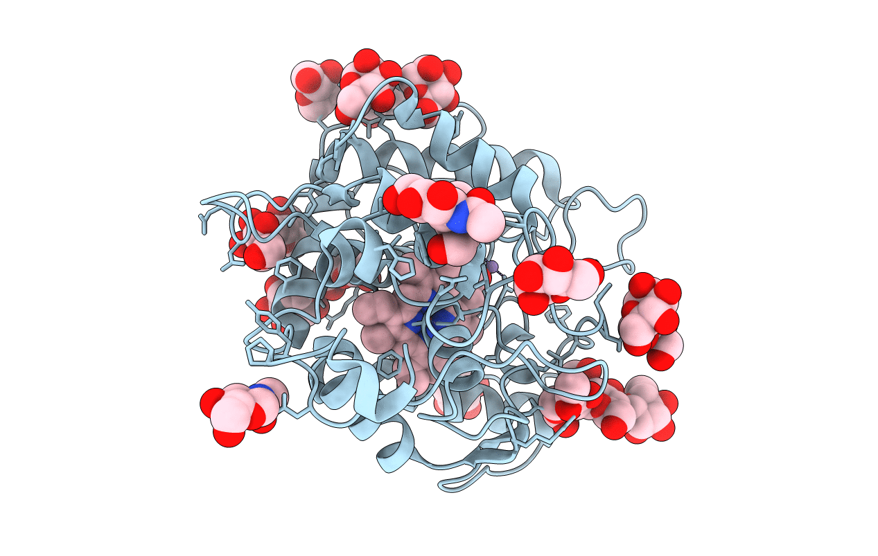
Structure of Chloroperoxidase Compound 0
Biological Source:
Source Organism:
Method Details:
Experimental Method:
Resolution:
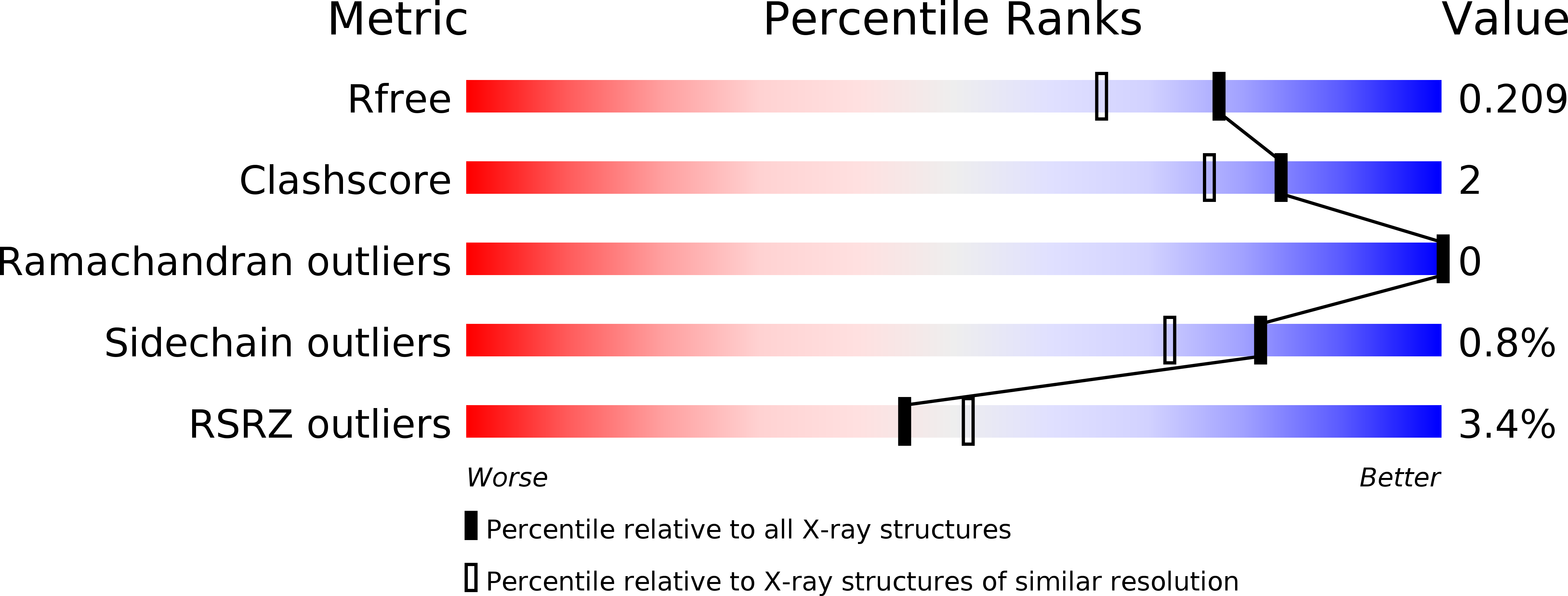
1.75 Å
R-Value Free:
0.21
R-Value Work:
0.19
R-Value Observed:
0.19
Space Group:
C 2 2 21
Macromolecular Entities
Structures with similar UniProt ID
Protein Blast
Polymer Type:polypeptide(L)
Molecule:CHLOROPEROXIDASE
Gene (Uniprot):CPO
Chain IDs:A
Chain Length:299
Number of Molecules:1
Biological Source:LEPTOXYPHIUM FUMAGO
Modified Residue
Compound ID Chain ID Parent Comp ID Details 2D Image
ASN A ASN GLYCOSYLATION SITE
PCA A GLU PYROGLUTAMIC ACID
SER A SER GLYCOSYLATION SITE
THR A THR GLYCOSYLATION SITE
Peptide-like Molecules
PRD_900111
Primary Citation
Structure and Quantum Chemical Characterization of Chloroperoxidase Compound 0, a Common Reaction Intermediate of Diverse Heme Enzymes.
Proc.Natl.Acad.Sci.USA 104 99 ? (2007)
PMID: 17190816 DOI: 10.1073/PNAS.0606285103

Abstact

We have determined the crystal structure of the chloroperoxidase (CPO) hydroperoxo reaction intermediate (CPO compound 0) at 1.75-A resolution. The intermediate was generated through controlled photoreduction of the CPO oxygen complex during x-ray data collection, which was monitored by recording of the crystal absorption spectra. Initially, the peroxo-anion species was formed and then protonated to yield compound 0. Quantum chemical calculations indicate that the peroxo-anion species is not stable and collapses instantaneously to compound 0. Compound 0 is present in the ferric low-spin doublet ground state and is characterized by a long O O bond length of 1.5 A and a Fe O bond distance of 1.8 A, which is also observed in the crystal structure.

Legend

Protein

Chemical

Disease

Primary Citation of related structures