Deposition Date
2006-08-23
Release Date
2006-10-11
Last Version Date
2024-05-08
Entry Detail
PDB ID:
2J3V
Keywords:
Title:
Crystal structure of the enzymatic component C2-I of the C2-toxin from Clostridium botulinum at pH 3.0
Biological Source:
Source Organism(s):
CLOSTRIDIUM BOTULINUM (Taxon ID: 1491)
Expression System(s):
Method Details:
Experimental Method:
Resolution:
2.11 Å
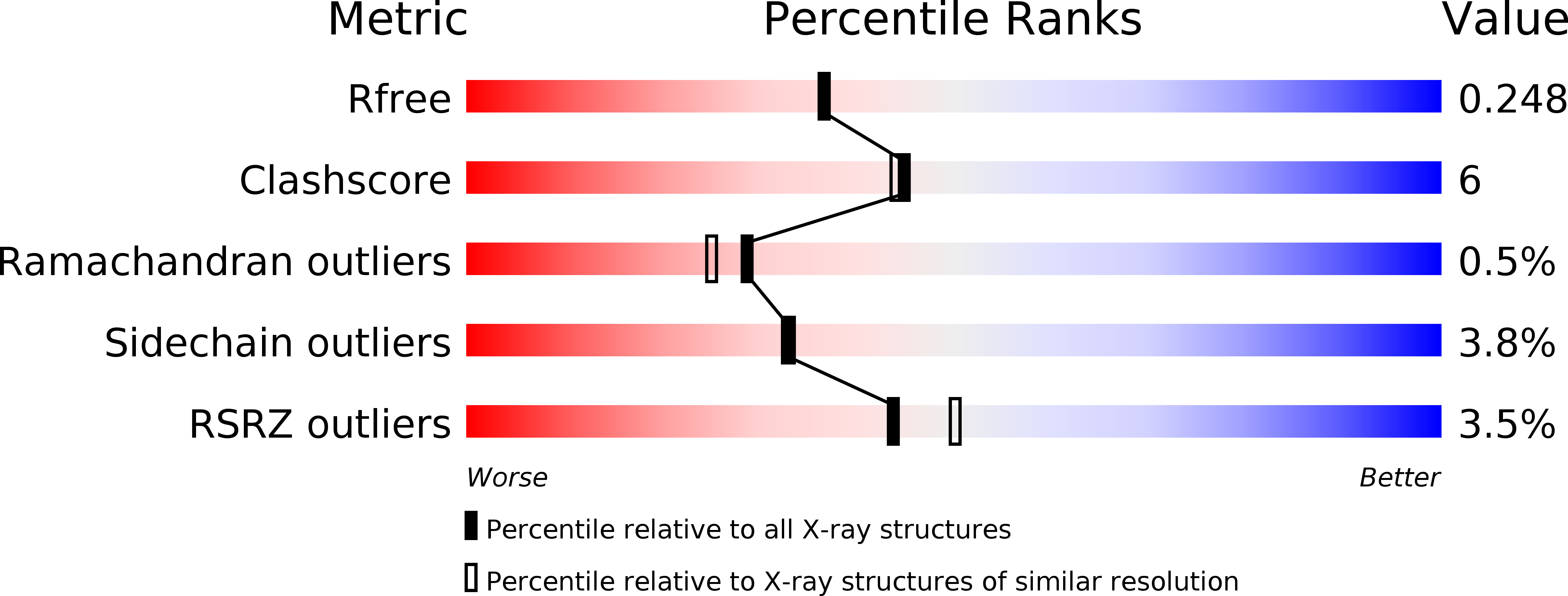
R-Value Free:
0.23
R-Value Work:
0.18
R-Value Observed:
0.19
Space Group:
P 63 2 2