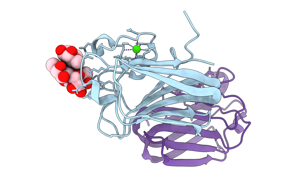
2J1T image
Deposition Date 2006-08-15
Release Date 2006-09-06
Last Version Date 2024-05-08
Entry Detail
PDB ID:
2J1T
Title:
Structure of a Streptococcus pneumoniae fucose binding module in complex with the Lewis Y antigen
Biological Source:
Source Organism:
Host Organism:
Method Details:
Experimental Method:
Resolution:
1.60 Å
R-Value Free:
0.20
R-Value Work:
0.14
R-Value Observed:
0.15
Space Group:
P 21 21 21
Macromolecular Entities
Structures with similar UniProt ID
Protein Blast
Polymer Type:polypeptide(L)
Molecule:FUCOLECTIN-RELATED PROTEIN
Chain IDs:A, B
Chain Length:151
Number of Molecules:2
Biological Source:STREPTOCOCCUS PNEUMONIAE
Ligand Molecules
Peptide-like Molecules
PRD_900054
Primary Citation
Blood Group Antigen Recognition by a Streptococcus Pneumoniae Virulence Factor
J.Biol.Chem. 281 35263 ? (2006)
PMID: 16987809 DOI: 10.1074/JBC.M607620200

Abstact

The Streptococcus pneumoniae fucose utilization operon includes a gene encoding a virulence factor that belongs to family 98 in the glycoside hydrolase classification. This protein contains a C-terminal triplet of fucose binding modules that have significant amino acid sequence identity with the Anguilla anguilla fucolectin. Functional studies of these fucose binding modules reveal binding to fucosylated oligosaccharides and suggest the importance of multivalent binding. The high resolution crystal structures of ligand bound forms of one fucose binding module uncovers the molecular basis of fucose, ABH blood group antigen, and Lewisy antigen binding. These studies are extended by fluorescence microscopy to show specific binding to mouse lung tissue. These modules define a new family of carbohydrate binding modules now classified as family 47.

Legend

Protein

Chemical

Disease

Primary Citation of related structures