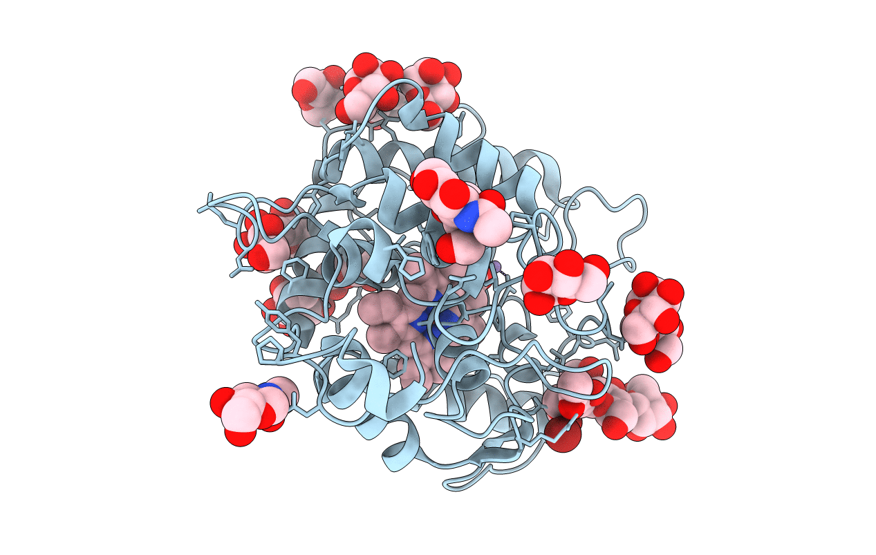
2J18 image
Deposition Date 2006-08-09
Release Date 2006-12-18
Last Version Date 2024-11-20
Entry Detail
PDB ID:
2J18
Keywords:
Title:
Chloroperoxidase mixture of ferric and ferrous states (low dose data set)
Biological Source:
Source Organism:
Host Organism:
Method Details:
Experimental Method:
Resolution:
1.75 Å
R-Value Free:
0.20
R-Value Work:
0.17
R-Value Observed:
0.17
Space Group:
C 2 2 21
Macromolecular Entities
Structures with similar UniProt ID
Protein Blast
Polymer Type:polypeptide(L)
Molecule:CHLOROPEROXIDASE
Gene (Uniprot):CPO
Chain IDs:A
Chain Length:299
Number of Molecules:1
Biological Source:CALDARIOMYCES FUMAGO
Modified Residue
Compound ID Chain ID Parent Comp ID Details 2D Image
ASN A ASN GLYCOSYLATION SITE
PCA A GLU PYROGLUTAMIC ACID
SER A SER GLYCOSYLATION SITE
THR A THR GLYCOSYLATION SITE
Peptide-like Molecules
PRD_900111
Primary Citation
Cryoradiolytic Reduction of Crystalline Heme Proteins: Analysis by Uv-Vis Spectroscopy and X-Ray Crystallography
J.Synchrotron Radiat. 14 11 ? (2007)
PMID: 17211068 DOI: 10.1107/S0909049506049806

Abstact

The X-ray crystallographic analysis of redox-active systems may be complicated by photoreduction. Although radiolytic reduction by the probing X-ray beam may be exploited to generate otherwise short-lived reaction intermediates of metalloproteins, it is generally an undesired feature. Here, the X-ray-induced reduction of the three heme proteins myoglobin, cytochrome P450cam and chloroperoxidase has been followed by on-line UV-Vis absorption spectroscopy. All three systems showed a very rapid reduction of the heme iron. In chloroperoxidase the change of the ionization state from ferric to ferrous heme is associated with a movement of the heme-coordinating water molecule. The influence of the energy of the incident X-ray photons and of the presence of scavengers on the apparent reduction rate of ferric myoglobin crystals was analyzed.

Legend

Protein

Chemical

Disease

Primary Citation of related structures