Deposition Date
2006-08-08
Release Date
2007-08-28
Last Version Date
2024-05-15
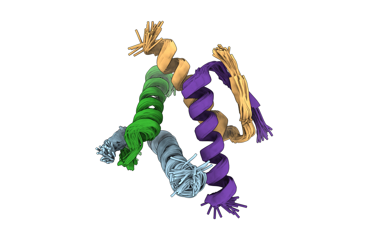
Method Details:
Experimental Method:
Conformers Calculated:
30
Conformers Submitted:
30
Selection Criteria:
TOTAL ENERGY