2IYJ image
Deposition Date 2006-07-18
Release Date 2007-07-24
Last Version Date 2023-12-13
Entry Detail
PDB ID:
2IYJ
Keywords:
Title:
Crystal structure of the N-terminal dimer domain of E.coli DsbC
Biological Source:
Source Organism:
ESCHERICHIA COLI (Taxon ID: 562)
Method Details:
Experimental Method:
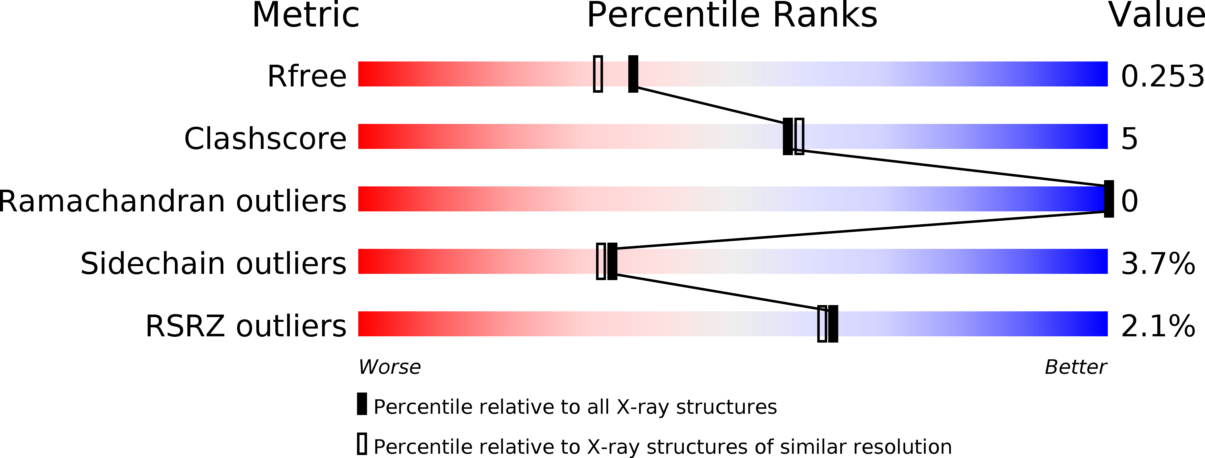
Resolution:
2.00 Å
R-Value Free:
0.25
R-Value Work:
0.22
R-Value Observed:
0.22
Space Group:
P 65 2 2
Macromolecular Entities
Structures with similar UniProt ID
Protein Blast
Polymer Type:polypeptide(L)
Molecule:THIOL DISULFIDE INTERCHANGE PROTEIN DSBC
Chain IDs:A, B
Chain Length:75
Number of Molecules:2
Biological Source:ESCHERICHIA COLI
Ligand Molecules
Primary Citation
Structures of Dimerization Domains of the Escherichia Coli Disulfide-Bond Isomerase Enzymes Dsbc and Dsbg.
Acta Crystallogr.,Sect.D 63 465 ? (2007)
PMID: 17372350 DOI: 10.1107/S0907444907003320

Abstact

DsbC and DsbG are periplasmic disulfide-bond isomerases, enzymes that facilitate the folding of secreted proteins with multiple disulfide bonds by catalyzing disulfide-bond rearrangement. Both enzymes also have in vitro chaperone activity. The crystal structures of these molecules are similar and both are V-shaped homodimeric modular structures. Each dimeric molecule contains two separate C-terminal thioredoxin-fold domains, joined by hinged helical "stalks" to a single N-terminal dimerization domain formed from the N-terminal 67 residues of each monomer. In this work, the crystal structures of the separate DsbC and DsbG dimerization domains have been determined at resolutions of 2.0 and 1.9 A, respectively. The two structures are both similar to the corresponding domains in the full-length molecules, showing that the dimerization domains fold independently of the catalytic portions of the full-length molecules. Localized structural differences between DsbC and DsbG were observed near the dimer interface and may be relevant to the different functions of the two enzymes.

Legend

Protein

Chemical

Disease

Primary Citation of related structures