2IY5 image
Deposition Date 2006-07-12
Release Date 2006-09-06
Last Version Date 2023-12-13
Entry Detail
PDB ID:
2IY5
Keywords:
Title:
PHENYLALANYL-TRNA SYNTHETASE FROM THERMUS THERMOPHILUS complexed with tRNA and a phenylalanyl-adenylate analog
Biological Source:
Source Organism:
Method Details:
Experimental Method:
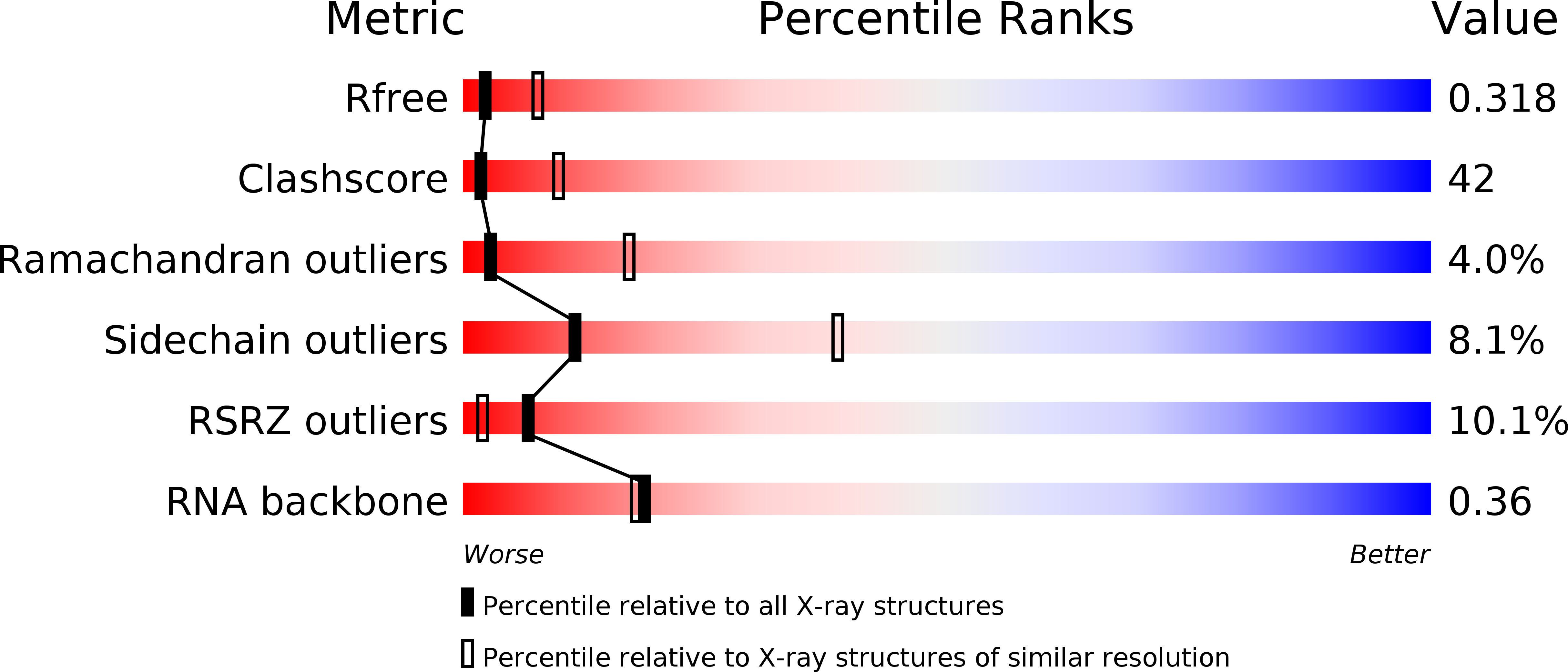
Resolution:
3.10 Å
R-Value Free:
0.29
R-Value Work:
0.23
R-Value Observed:
0.23
Space Group:
P 32 2 1
Macromolecular Entities
Structures with similar UniProt ID
Protein Blast
Polymer Type:polypeptide(L)
Molecule:PHENYLALANYL-TRNA SYNTHETASE ALPHA CHAIN
Chain IDs:A
Chain Length:350
Number of Molecules:1
Biological Source:THERMUS THERMOPHILUS
Structures with similar UniProt ID
Protein Blast
Polymer Type:polypeptide(L)
Molecule:PHENYLALANYL-TRNA SYNTHETASE BETA CHAIN
Chain IDs:B
Chain Length:785
Number of Molecules:1
Biological Source:THERMUS THERMOPHILUS
Polymer Type:polyribonucleotide
Molecule:TRNAPHE
Chain IDs:C (auth: T)
Chain Length:76
Number of Molecules:1
Biological Source:THERMUS THERMOPHILUS
Primary Citation
The crystal structure of the ternary complex of phenylalanyl-tRNA synthetase with tRNAPhe and a phenylalanyl-adenylate analogue reveals a conformational switch of the CCA end.
Biochemistry 45 10572 10583 (2006)
PMID: 16939209 DOI: 10.1021/bi060491l

Abstact

The crystal structure of the ternary complex of (alphabeta)(2) heterotetrameric phenylalanyl-tRNA synthetase (PheRS) from Thermus thermophilus with cognate tRNA(Phe) and a nonhydrolyzable phenylalanyl-adenylate analogue (PheOH-AMP) has been determined at 3.1 A resolution. It reveals conformational changes in tRNA(Phe) induced by the PheOH-AMP binding. The single-stranded 3' end exhibits a hairpin conformation in contrast to the partial unwinding observed previously in the binary PheRS.tRNA(Phe) complex. The CCA end orientation is stabilized by extensive base-specific interactions of A76 and C75 with the protein and by intra-RNA interactions of A73 with adjacent nucleotides. The 4-amino group of the "bulged out" C75 is trapped by two negatively charged residues of the beta subunit (Glubeta31 and Aspbeta33), highly conserved in eubacterial PheRSs. The position of the A76 base is stabilized by interactions with Hisalpha212 of motif 2 (universally conserved in PheRSs) and class II-invariant Argalpha321 of motif 3. Important conformational changes induced by the binding of tRNA(Phe) and PheOH-AMP are observed in the catalytic domain: the motif 2 loop and a "helical" loop (residues 139-152 of the alpha subunit) undergo coordinated displacement; Metalpha148 of the helical loop adopts a conformation preventing the 2'-OH group of A76 from approaching the alpha-carbonyl carbon of PheOH-AMP. The unfavorable position of the terminal ribose stems from the absence of the alpha-carbonyl oxygen in the analogue. Our data suggest that the idiosyncratic feature of PheRS, which aminoacylates the 2'-OH group of the terminal ribose, is dictated by the system-specific topology of the CCA end-binding site.

Legend

Protein

Chemical

Disease

Primary Citation of related structures