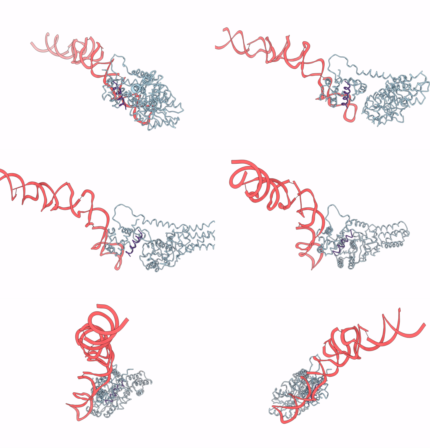
2IY3 image
Deposition Date 2006-07-12
Release Date 2006-11-02
Last Version Date 2024-05-08
Entry Detail
PDB ID:
2IY3
Keywords:
Title:
Structure of the E. Coli Signal Regognition Particle
Biological Source:
Source Organism:
Host Organism:
Method Details:
Experimental Method:
Resolution:
16.00 Å
Aggregation State:
PARTICLE
Reconstruction Method:
SINGLE PARTICLE
Macromolecular Entities
Protein Blast
Polymer Type:polypeptide(L)
Molecule:Signal recognition particle protein,Signal recognition particle 54 kDa protein
Gene (Uniprot):ffh
Chain IDs:A
Chain Length:432
Number of Molecules:1
Biological Source:Thermus aquaticus, Sulfolobus solfataricus
Polymer Type:polyribonucleotide
Molecule:4.5S RNA
Chain IDs:B
Chain Length:110
Number of Molecules:1
Biological Source:Escherichia coli
Polymer Type:polypeptide(L)
Molecule:SIGNAL SEQUENCE
Chain IDs:C
Chain Length:17
Number of Molecules:1
Biological Source: synthetic construct
Ligand Molecules
Primary Citation
Structure of the E. Coli Signal Recognition Particle Bound to a Translating Ribosome
Nature 444 503 ? (2006)
PMID: 17086205 DOI: 10.1038/NATURE05182

Abstact

The prokaryotic signal recognition particle (SRP) targets membrane proteins into the inner membrane. It binds translating ribosomes and screens the emerging nascent chain for a hydrophobic signal sequence, such as the transmembrane helix of inner membrane proteins. If such a sequence emerges, the SRP binds tightly, allowing the SRP receptor to lock on. This assembly delivers the ribosome-nascent chain complex to the protein translocation machinery in the membrane. Using cryo-electron microscopy and single-particle reconstruction, we obtained a 16 A structure of the Escherichia coli SRP in complex with a translating E. coli ribosome containing a nascent chain with a transmembrane helix anchor. We also obtained structural information on the SRP bound to an empty E. coli ribosome. The latter might share characteristics with a scanning SRP complex, whereas the former represents the next step: the targeting complex ready for receptor binding. High-resolution structures of the bacterial ribosome and of the bacterial SRP components are available, and their fitting explains our electron microscopic density. The structures reveal the regions that are involved in complex formation, provide insight into the conformation of the SRP on the ribosome and indicate the conformational changes that accompany high-affinity SRP binding to ribosome nascent chain complexes upon recognition of the signal sequence.

Legend

Protein

Chemical

Disease

Primary Citation of related structures