2IX1 image
Deposition Date 2006-07-05
Release Date 2006-10-05
Last Version Date 2024-05-08
Entry Detail
PDB ID:
2IX1
Keywords:
Title:
RNase II D209N mutant
Biological Source:
Source Organism:
Host Organism:
Method Details:
Experimental Method:
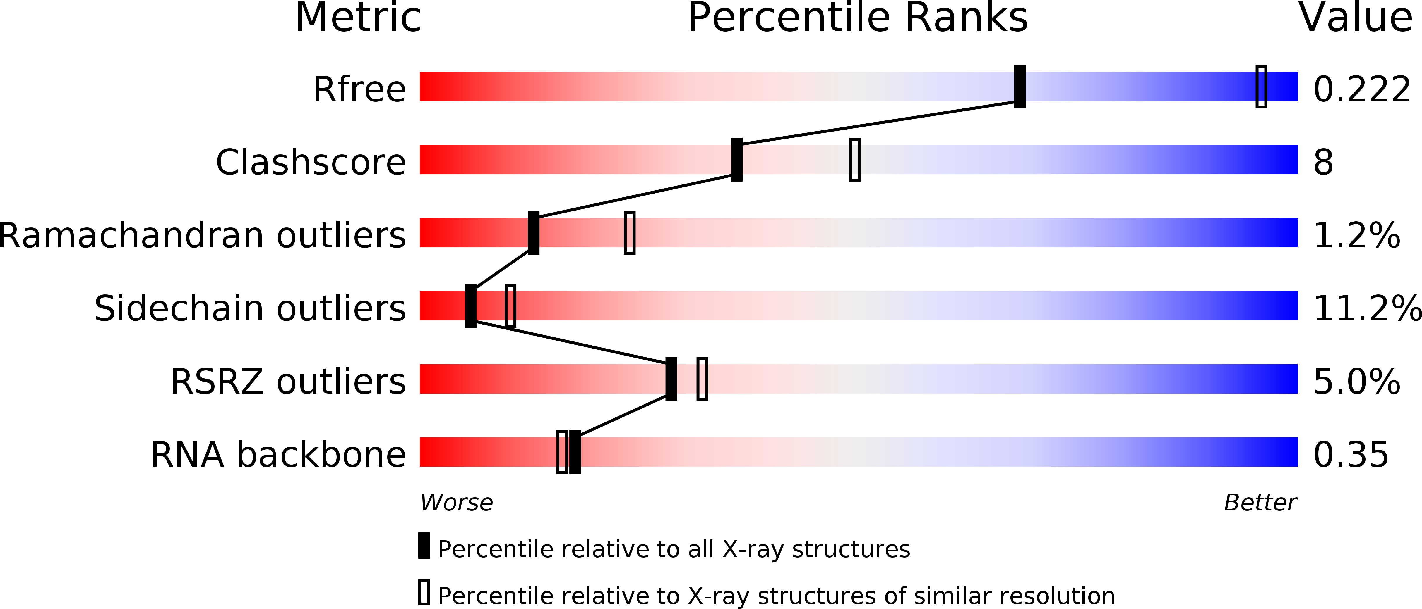
Resolution:
2.74 Å
R-Value Free:
0.22
R-Value Work:
0.18
R-Value Observed:
0.18
Space Group:
P 65
Macromolecular Entities
Polymer Type:polypeptide(L)
Molecule:EXORIBONUCLEASE 2
Gene (Uniprot):rnb
Mutations:YES
Chain IDs:A
Chain Length:664
Number of Molecules:1
Biological Source:ESCHERICHIA COLI
Polymer Type:polyribonucleotide
Molecule:5'-D(*AP*AP*AP*AP*AP*AP*AP*AP*AP*AP *AP*AP*A)-3'
Chain IDs:B
Chain Length:13
Number of Molecules:1
Biological Source:ESCHERICHIA COLI BL21(DE3)
Ligand Molecules
Primary Citation

Abstact

RNA degradation is a determining factor in the control of gene expression. The maturation, turnover and quality control of RNA is performed by many different classes of ribonucleases. Ribonuclease II (RNase II) is a major exoribonuclease that intervenes in all of these fundamental processes; it can act independently or as a component of the exosome, an essential RNA-degrading multiprotein complex. RNase II-like enzymes are found in all three kingdoms of life, but there are no structural data for any of the proteins of this family. Here we report the X-ray crystallographic structures of both the ligand-free (at 2.44 A resolution) and RNA-bound (at 2.74 A resolution) forms of Escherichia coli RNase II. In contrast to sequence predictions, the structures show that RNase II is organized into four domains: two cold-shock domains, one RNB catalytic domain, which has an unprecedented alphabeta-fold, and one S1 domain. The enzyme establishes contacts with RNA in two distinct regions, the 'anchor' and the 'catalytic' regions, which act synergistically to provide catalysis. The active site is buried within the RNB catalytic domain, in a pocket formed by four conserved sequence motifs. The structure shows that the catalytic pocket is only accessible to single-stranded RNA, and explains the specificity for RNA versus DNA cleavage. It also explains the dynamic mechanism of RNA degradation by providing the structural basis for RNA translocation and enzyme processivity. We propose a reaction mechanism for exonucleolytic RNA degradation involving key conserved residues. Our three-dimensional model corroborates all existing biochemical data for RNase II, and elucidates the general basis for RNA degradation. Moreover, it reveals important structural features that can be extrapolated to other members of this family.

Legend

Protein

Chemical

Disease

Primary Citation of related structures