2IWW image
Deposition Date 2006-07-05
Release Date 2006-08-14
Last Version Date 2024-05-08
Entry Detail
PDB ID:
2IWW
Keywords:
Title:
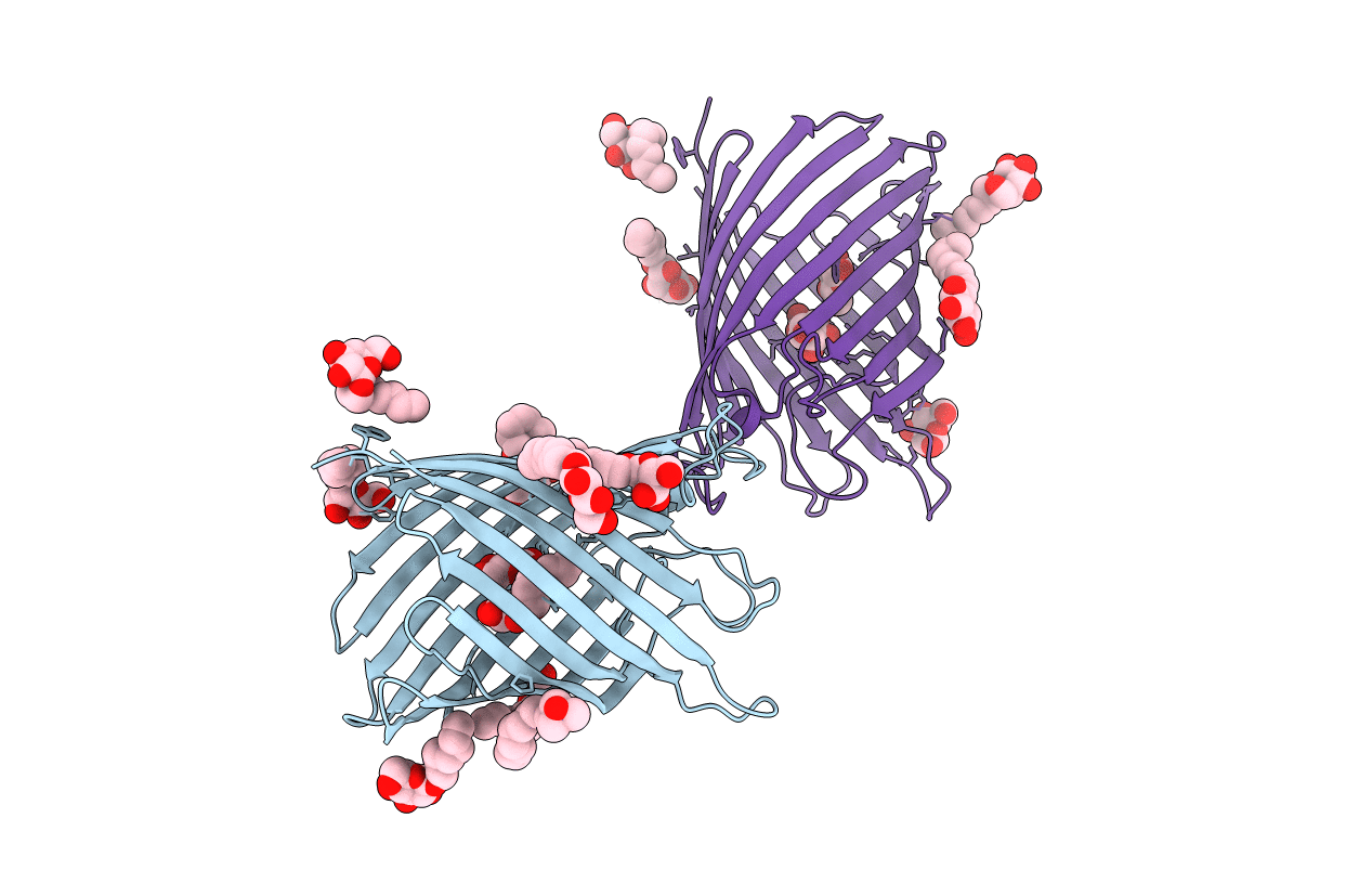
Structure of the monomeric outer membrane porin OmpG in the open and closed conformation
Biological Source:
Source Organism:
Host Organism:
Method Details:
Experimental Method:
Resolution:
2.70 Å
R-Value Free:
0.30
R-Value Work:
0.24
R-Value Observed:
0.24
Space Group:
P 21 21 21
Macromolecular Entities
Polymer Type:polypeptide(L)
Molecule:OUTER MEMBRANE PROTEIN G
Gene (Uniprot):ompG
Chain IDs:A, B
Chain Length:281
Number of Molecules:2
Biological Source:ESCHERICHIA COLI
Primary Citation
Structure of the Monomeric Outer-Membrane Porin Ompg in the Open and Closed Conformation.
Embo J. 25 3702 ? (2006)
PMID: 16888630 DOI: 10.1038/SJ.EMBOJ.7601237

Abstact

OmpG, a monomeric pore-forming protein from Escherichia coli outer membranes, was refolded from inclusion bodies and crystallized in two different conformations. The OmpG channel is a 14-stranded beta-barrel, with short periplasmic turns and seven extracellular loops. Crystals grown at neutral pH show the channel in the open state at 2.3 A resolution. In the 2.7 A structure of crystals grown at pH 5.6, the pore is blocked by loop 6, which folds across the channel. The rearrangement of loop 6 appears to be triggered by a pair of histidine residues, which repel one another at acidic pH, resulting in the breakage of neighbouring H-bonds and a lengthening of loop 6 from 10 to 17 residues. A total of 151 ordered LDAO detergent molecules were found in the 2.3 A structure, mostly on the hydrophobic outer surface of OmpG, mimicking the outer membrane lipid bilayer, with three LDAO molecules in the open pore. In the 2.7 A structure, OmpG binds one OG and one glucose molecule as sugar substrates in the closed pore.

Legend

Protein

Chemical

Disease

Primary Citation of related structures