2IWL image
Deposition Date 2006-07-01
Release Date 2006-10-18
Last Version Date 2024-05-08
Entry Detail
PDB ID:
2IWL
Keywords:
Title:
Structure of the PX Domain of Phosphoinositide 3-Kinase-C2alpha
Biological Source:
Source Organism:
HOMO SAPIENS (Taxon ID: 9606)
Method Details:
Experimental Method:
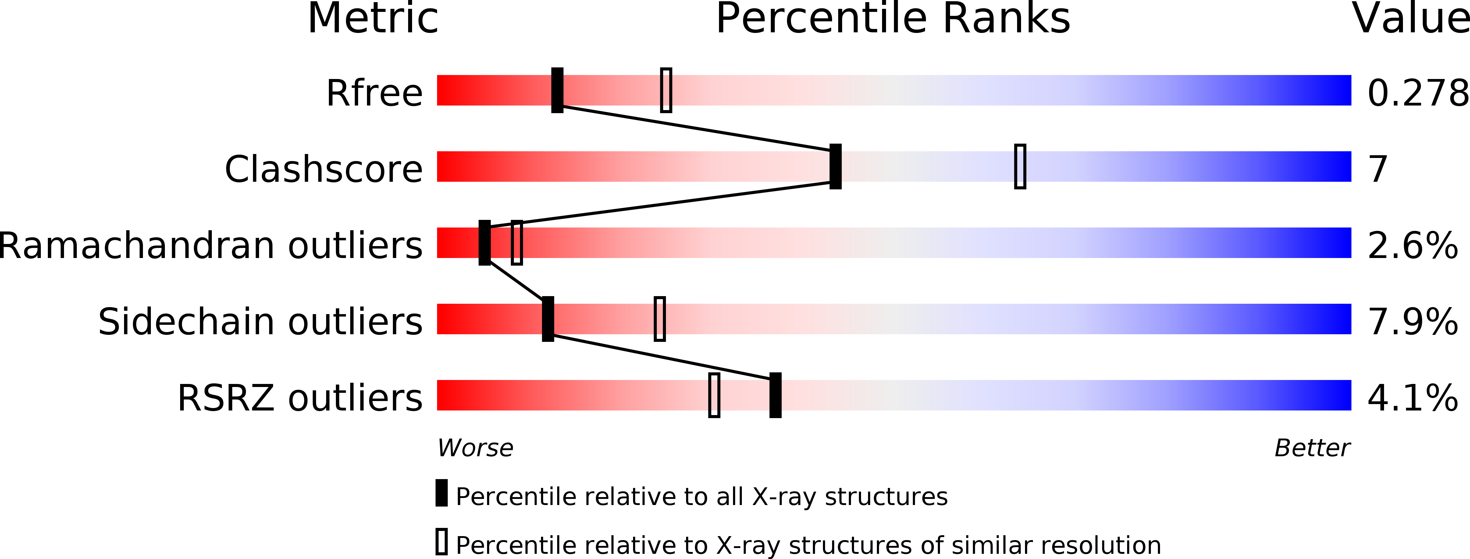
Resolution:
2.60 Å
R-Value Free:
0.29
R-Value Work:
0.24
R-Value Observed:
0.25
Space Group:
P 4 3 2
Macromolecular Entities
Polymer Type:polypeptide(L)
Molecule:PHOSPHATIDYLINOSITOL-4-PHOSPHATE 3-KINASE C2 DOMAIN-CONTAINING ALPHA POLYPEPTIDE
Gene (Uniprot):PIK3C2A
Chain IDs:A (auth: X)
Chain Length:140
Number of Molecules:1
Biological Source:HOMO SAPIENS
Ligand Molecules
Primary Citation
Structural and Membrane Binding Analysis of the Phox Homology Domain of Phosphoinositide 3-Kinase- C2{Alpha}.
J.Biol.Chem. 281 39396 ? (2006)
PMID: 17038310 DOI: 10.1074/JBC.M607079200

Abstact

Phox homology (PX) domains, which have been identified in a variety of proteins involved in cell signaling and membrane trafficking, have been shown to interact with phosphoinositides (PIs) with different affinities and specificities. To elucidate the structural origin of diverse PI specificities of PX domains, we determined the crystal structure of the PX domain from phosphoinositide 3-kinase C2alpha (PI3K-C2alpha), which binds phosphatidylinositol 4,5-bisphosphate (PtdIns(4,5)P(2)). To delineate the mechanism by which this PX domain interacts with membranes, we measured the membrane binding of the wild type domain and mutants by surface plasmon resonance and monolayer techniques. This PX domain contains a signature PI-binding site that is optimized for PtdIns(4,5)P(2) binding. The membrane binding of the PX domain is initiated by nonspecific electrostatic interactions followed by the membrane penetration of hydrophobic residues. Membrane penetration is specifically enhanced by PtdIns(4,5)P(2). Furthermore, the PX domain displayed significantly higher PtdIns(4,5)P(2) membrane affinity and specificity when compared with the PI3K-C2alpha C2 domain, demonstrating that high affinity PtdIns(4,5)P(2) binding was facilitated by the PX domain in full-length PI3K-C2alpha. Together, these studies provide new structural insight into the diverse PI specificities of PX domains and elucidate the mechanism by which the PI3K-C2alpha PX domain interacts with PtdIns(4,5)P(2)-containing membranes and thereby mediates the membrane recruitment of PI3K-C2alpha.

Legend

Protein

Chemical

Disease

Primary Citation of related structures