
Deposition Date
2006-06-20
Release Date
2007-02-13
Last Version Date
2024-05-15
Entry Detail
PDB ID:
2IVW
Keywords:
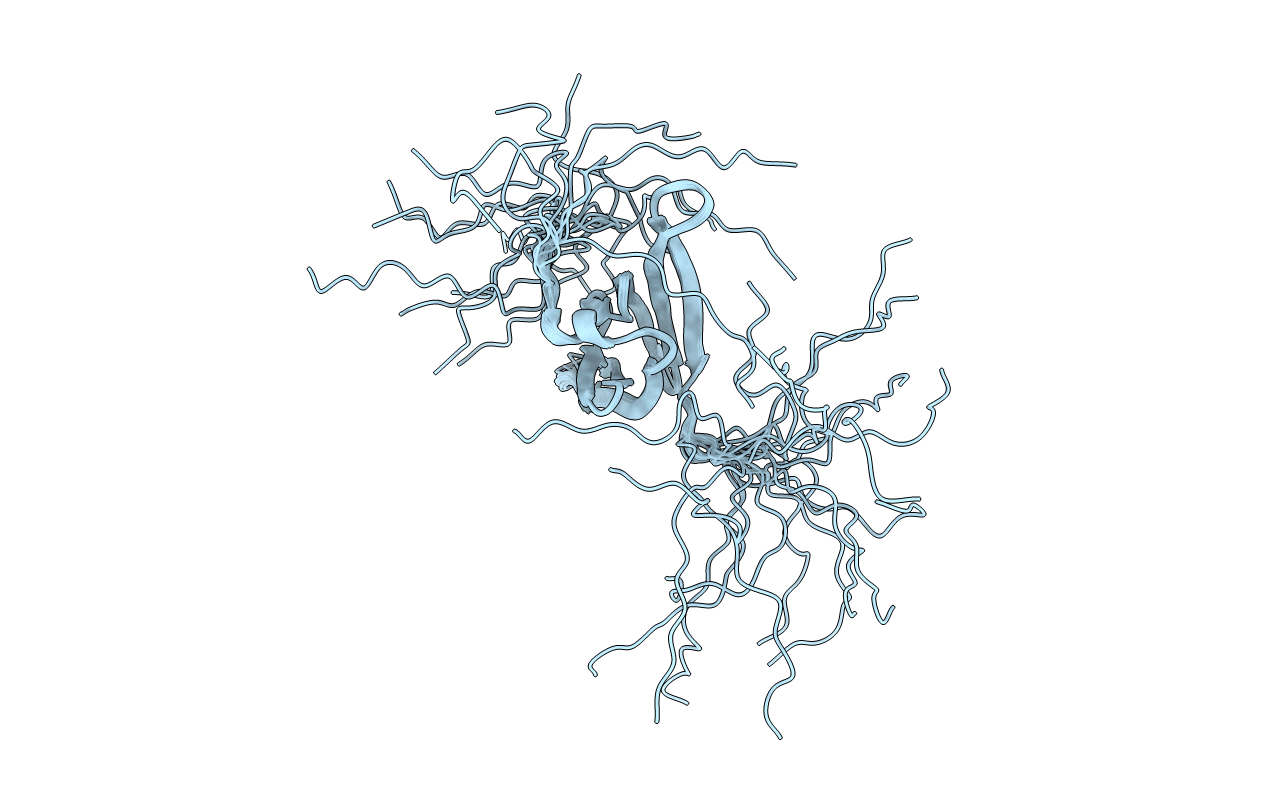
Title:
The solution structure of a domain from the Neisseria meningitidis PilP pilot protein.
Biological Source:
Source Organism(s):
NEISSERIA MENINGITIDIS (Taxon ID: 122587)
Expression System(s):
Method Details:
Experimental Method:
Conformers Calculated:
100
Conformers Submitted:
20
Selection Criteria:
LOWEST TARGET FUNCTION
