2IUI image
Deposition Date 2006-06-03
Release Date 2006-06-06
Last Version Date 2024-11-20
Entry Detail
PDB ID:
2IUI
Keywords:
Title:
Crystal structure of the PI3-kinase p85 N-terminal SH2 domain in complex with PDGFR phosphotyrosyl peptide
Biological Source:
Source Organism:
Homo sapiens (Taxon ID: 9606)
Method Details:
Experimental Method:
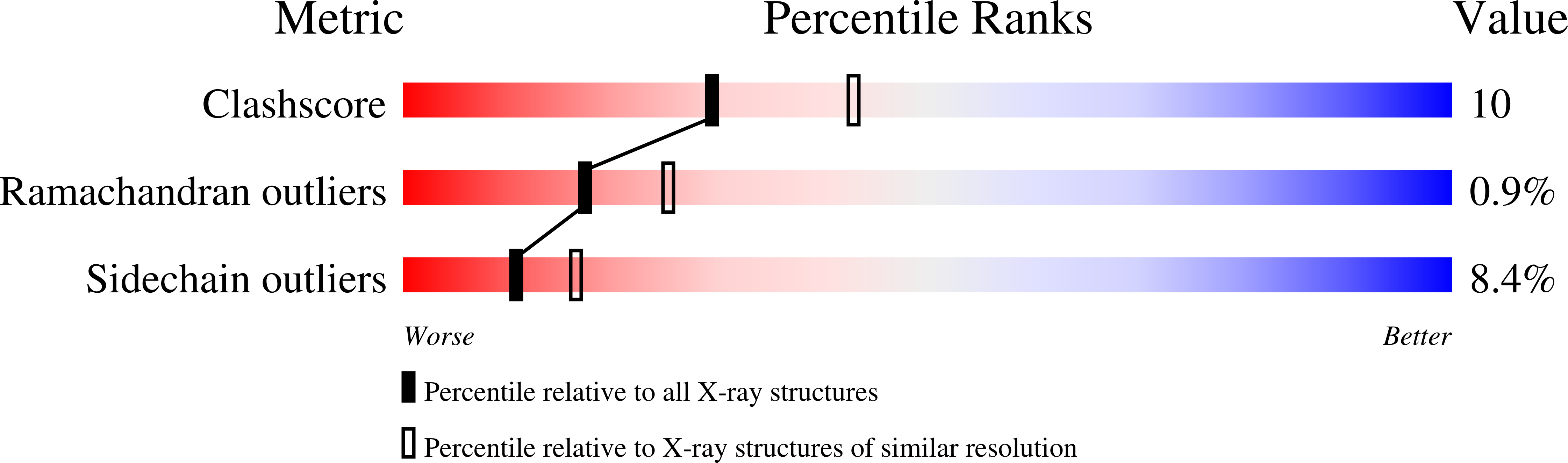
Resolution:
2.40 Å
R-Value Free:
0.24
R-Value Work:
0.17
R-Value Observed:
0.17
Space Group:
P 21 21 21
Macromolecular Entities
Polymer Type:polypeptide(L)
Molecule:Phosphatidylinositol 3-kinase regulatory subunit alpha
Gene (Uniprot):PIK3R1
Chain IDs:A, B
Chain Length:120
Number of Molecules:2
Biological Source:Homo sapiens
Polymer Type:polypeptide(L)
Molecule:Platelet-derived growth factor receptor beta
Gene (Uniprot):PDGFRB
Chain IDs:C, D
Chain Length:11
Number of Molecules:2
Biological Source:Homo sapiens
Modified Residue
Compound ID Chain ID Parent Comp ID Details 2D Image
PTR C TYR modified residue
Primary Citation
Crystal Structure of the Pi 3-Kinase P85 Amino- Terminal Sh2 Domain and its Phosphopeptide Complexes
Nat.Struct.Biol. 3 364 ? (1996)
PMID: 8599763 DOI: 10.1038/NSB0496-364

Abstact

Crystal structures of the amino-terminal SH2 domain of the p85alpha subunit of phosphatidylinositol (PI) 3-kinase, alone and in complex with phosphopeptides bearing pTyr-Met/Val-Xaa-Met motifs, show that phosphopeptides bind in the two-pronged manner seen in high-affinity Lck and Src SH2 complexes, with conserved interactions between the domain and the peptide segment from phosphotyrosine to Met+3. Peptide binding requires the rearrangement of a tyrosyl side chain in the BG loop to create the hydrophobic Met+3 binding pocket. The structures suggest a mechanism for the biological specificity exhibited by PI 3-kinase in its interactions with phosphoprotein partners.

Legend

Protein

Chemical

Disease

Primary Citation of related structures